Copyright
©The Author(s) 2021.
World J Clin Cases. Apr 16, 2021; 9(11): 2446-2457
Published online Apr 16, 2021. doi: 10.12998/wjcc.v9.i11.2446
Published online Apr 16, 2021. doi: 10.12998/wjcc.v9.i11.2446
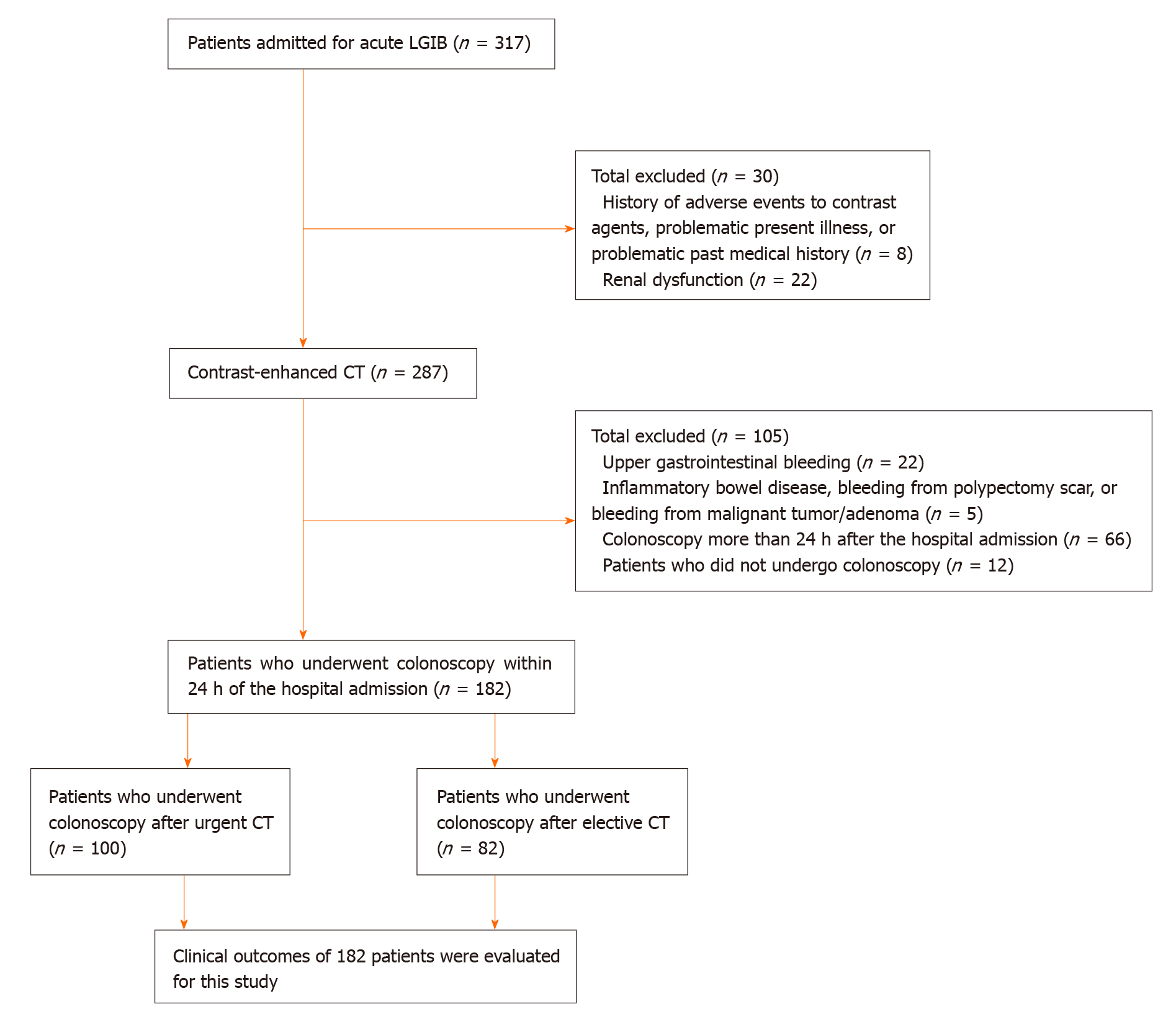
Figure 1 Flowchart for identification of patients with colonic diverticular bleeding.
LGIB: Lower gastrointestinal bleeding; CT: Computed tomography.
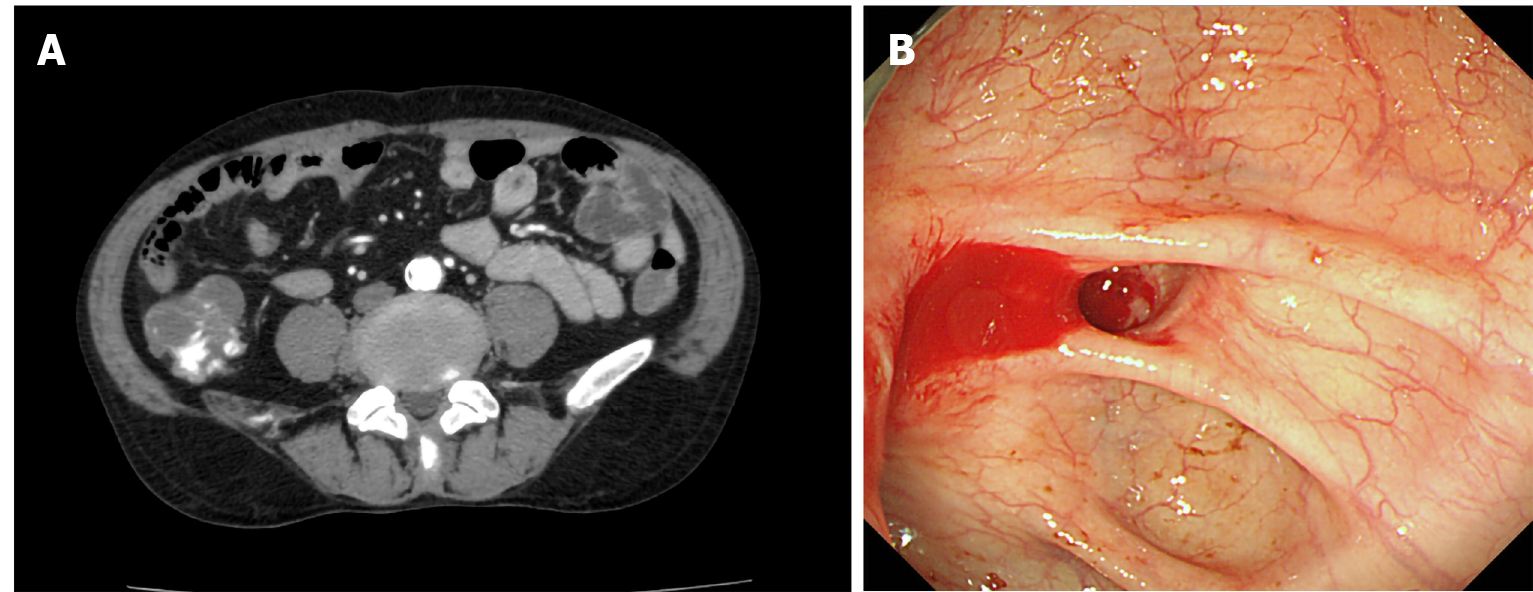
Figure 2 Image of stigmata of recent hemorrhage (active bleeding) identified on an extravasation-positive image by computed tomography.
A: An extravasation-positive image in the colonic lumen by contrast-enhanced computed tomography; B: Endoscopic view of active bleeding from a diverticulum.
- Citation: Ochi M, Kamoshida T, Hamano Y, Ohkawara A, Ohkawara H, Kakinoki N, Yamaguchi Y, Hirai S, Yanaka A. Early colonoscopy and urgent contrast enhanced computed tomography for colonic diverticular bleeding reduces risk of rebleeding. World J Clin Cases 2021; 9(11): 2446-2457
- URL: https://www.wjgnet.com/2307-8960/full/v9/i11/2446.htm
- DOI: https://dx.doi.org/10.12998/wjcc.v9.i11.2446


