Copyright
©The Author(s) 2021.
World J Clin Cases. Apr 6, 2021; 9(10): 2409-2418
Published online Apr 6, 2021. doi: 10.12998/wjcc.v9.i10.2409
Published online Apr 6, 2021. doi: 10.12998/wjcc.v9.i10.2409
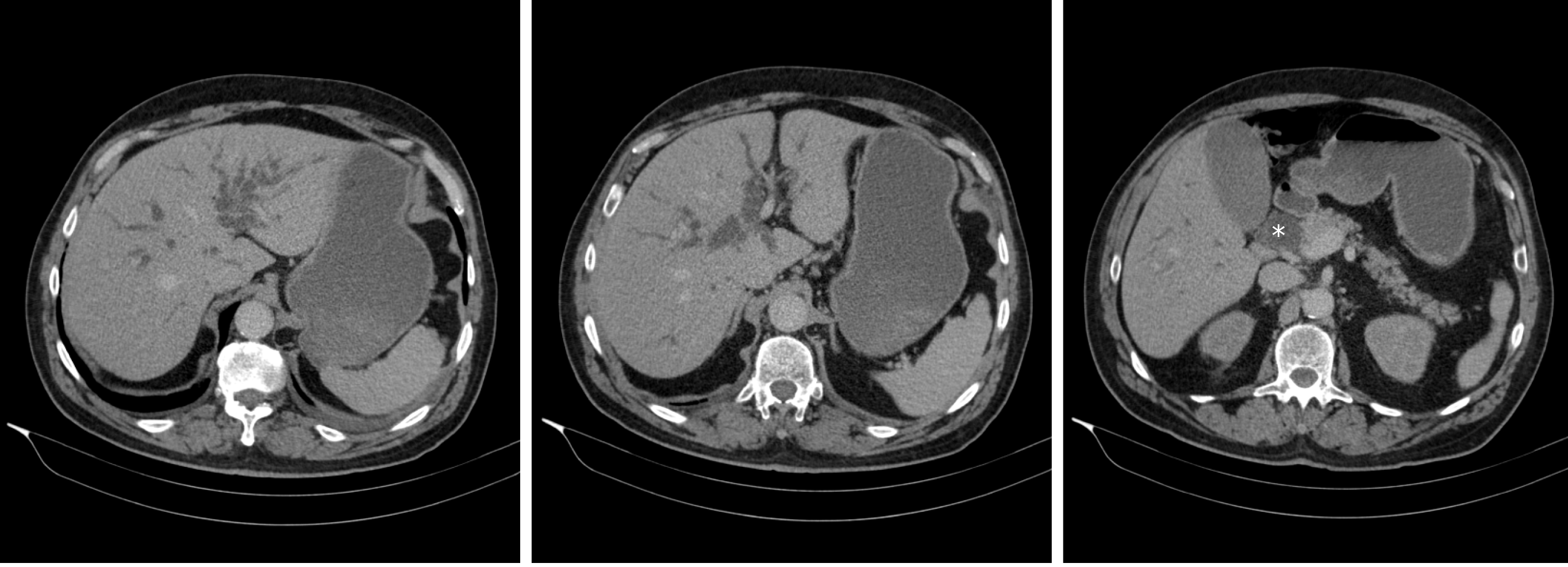
Figure 1 Abdominal computed tomography scan showing intra- and extrahepatic bile duct dilatation.
Dilated distal part of the common bile duct up to 2.6 cm is seen (asterisk) with no focal lesions identified.
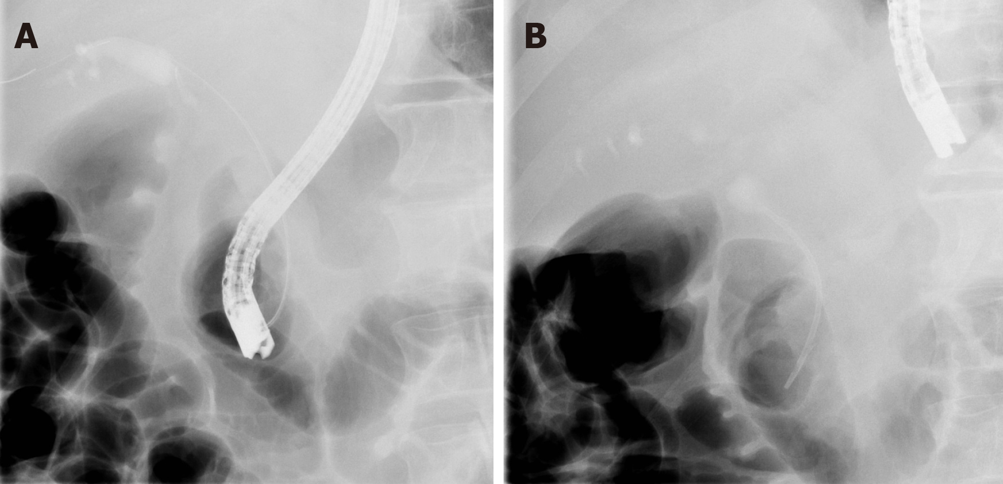
Figure 2 Endoscopic retrograde cholangiopancreatography.
A: Distal bile duct stenosis is seen with prestenotic dilatation; B: Biliary stent was inserted to overcome the stenosis.
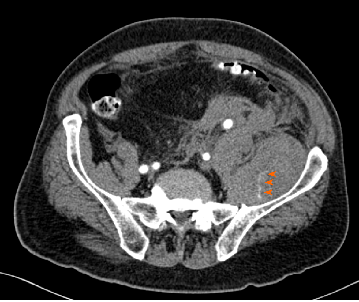
Figure 3 Computed tomography angiography of the abdomen showing retroperitoneal haemorrhage and haematoma in the left iliacus muscle with active contrast extravasation in venous phase (arrow heads).
- Citation: Krašek V, Kotnik A, Zavrtanik H, Klen J, Zver S. Acquired haemophilia in patients with malignant disease: A case report. World J Clin Cases 2021; 9(10): 2409-2418
- URL: https://www.wjgnet.com/2307-8960/full/v9/i10/2409.htm
- DOI: https://dx.doi.org/10.12998/wjcc.v9.i10.2409


