Copyright
©The Author(s) 2021.
World J Clin Cases. Apr 6, 2021; 9(10): 2386-2393
Published online Apr 6, 2021. doi: 10.12998/wjcc.v9.i10.2386
Published online Apr 6, 2021. doi: 10.12998/wjcc.v9.i10.2386
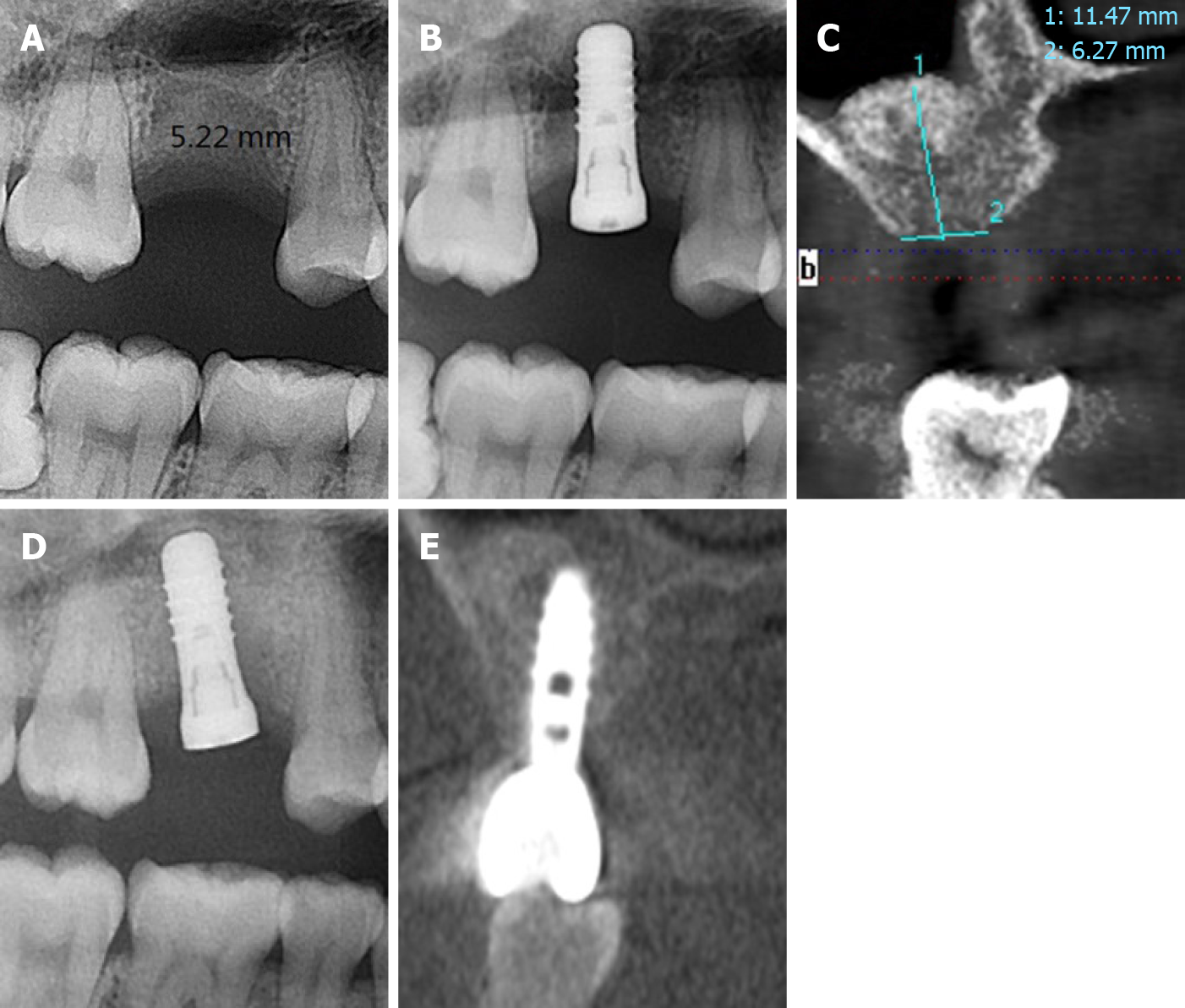
Figure 1 X-ray images of case 1.
A: In the preoperative X-ray image of the first-stage surgery, the residual bone height was 5.22 mm; B: In the postoperative X-ray image of the first-stage surgery, we placed a 4.8 mm-wide and 10 mm-long implant (Straumann); C: In the preoperative X-ray image of the second-stage surgery, the residual bone height was 11.47 mm; D: In the postoperative X-ray image of the second-stage surgery, we placed a 4.8 mm-wide and 10 mm-long implant (Straumann); E: The X-ray image after prosthetic rehabilitation.
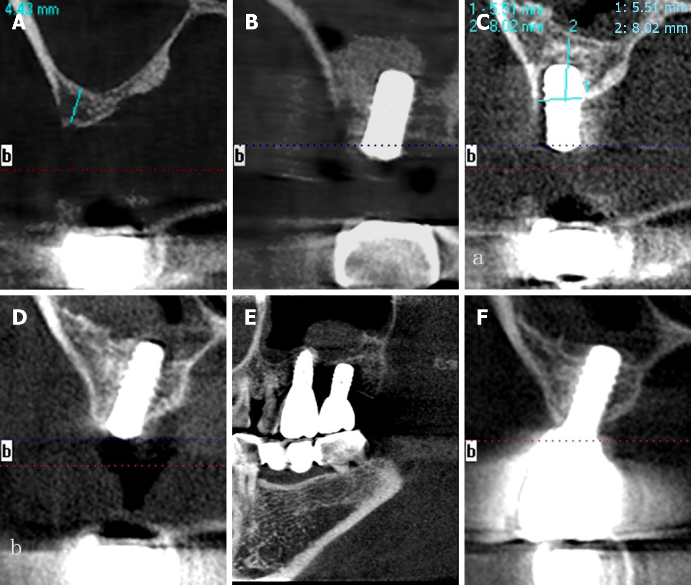
Figure 2 X-ray images of case 2.
A: In the preoperative X-ray image of the first-stage surgery, the residual bone height was 4.43 mm; B: In the postoperative X-ray image of the first-stage surgery, we placed a 4.8 mm-wide and 10 mm-long implant (Straumann); C: In the preoperative X-ray image of the second-stage surgery, the residual bone height was 8.02 mm; D: In the postoperative X-ray image of the second-stage surgery, we placed a 4.8 mm-wide and 10 mm-long implant (Straumann); E: The X-ray image after prosthetic rehabilitation; F: The x-ray image after prosthetic rehabilitation.
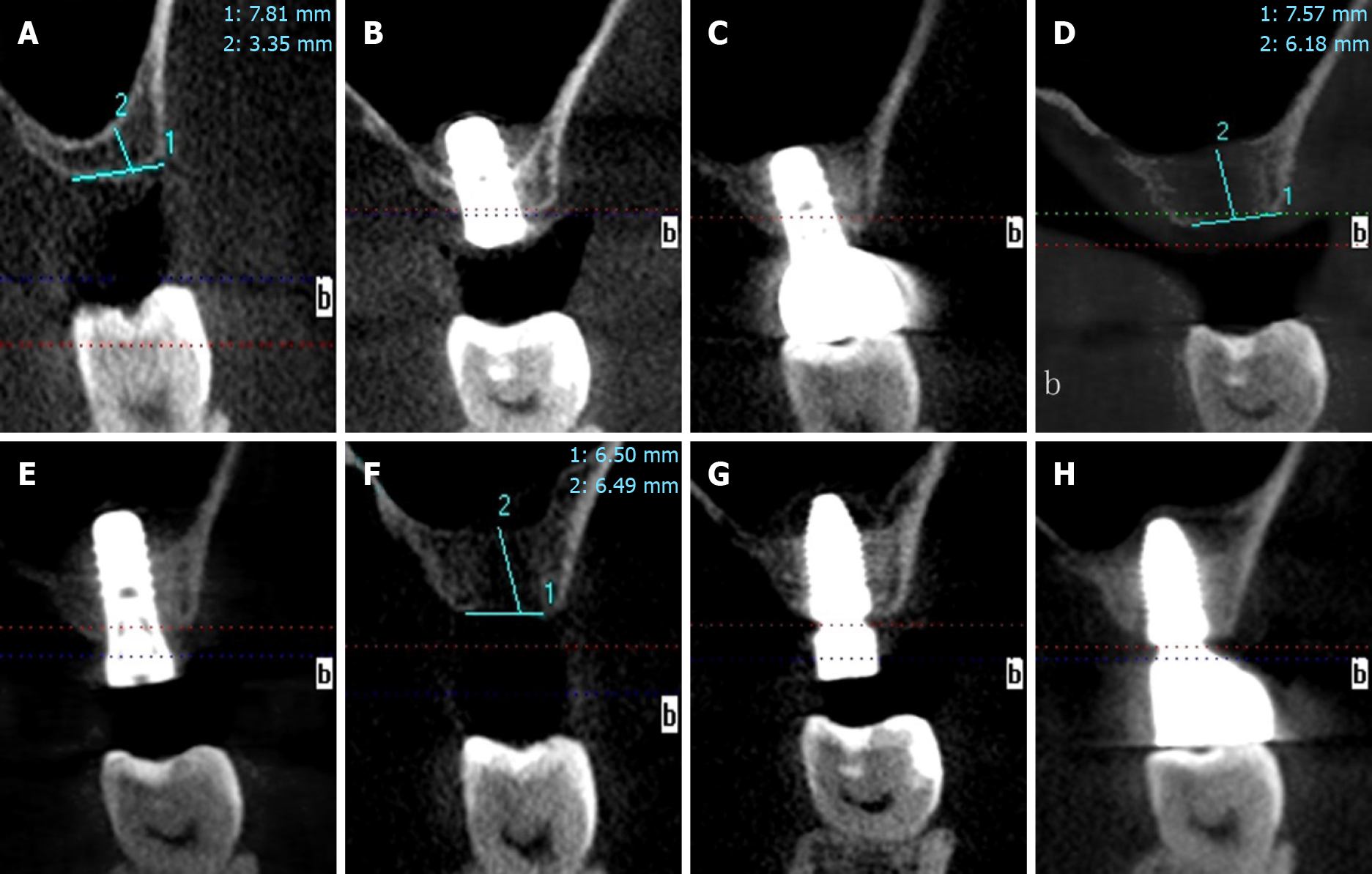
Figure 3 X-ray images of case 3.
A: In the preoperative X-ray image of the first-stage surgery, the residual bone height was 3.35 mm; B: In the postoperative X-ray image of the first-stage surgery, we placed a 4.8 mm-wide and 8 mm-long implant (Straumann); C: The X-ray image after prosthetic rehabilitation; D: In the preoperative X-ray image of the second-stage surgery, the residual bone height was 6.18 mm; E: In the postoperative X-ray image of the second-stage surgery, we placed a 4.8 mm-wide and 10 mm-long implant (Straumann), the implant was found to be mobile after 4 months; F: In the preoperative X-ray image of the third-stage surgery, the residual bone height was 6.49 mm; G: In the postoperative X-ray image of the third-stage surgery, we placed a 5.0 mm-wide and 10 mm-long implant (Nobel); H: The X-ray image after prosthetic rehabilitation.
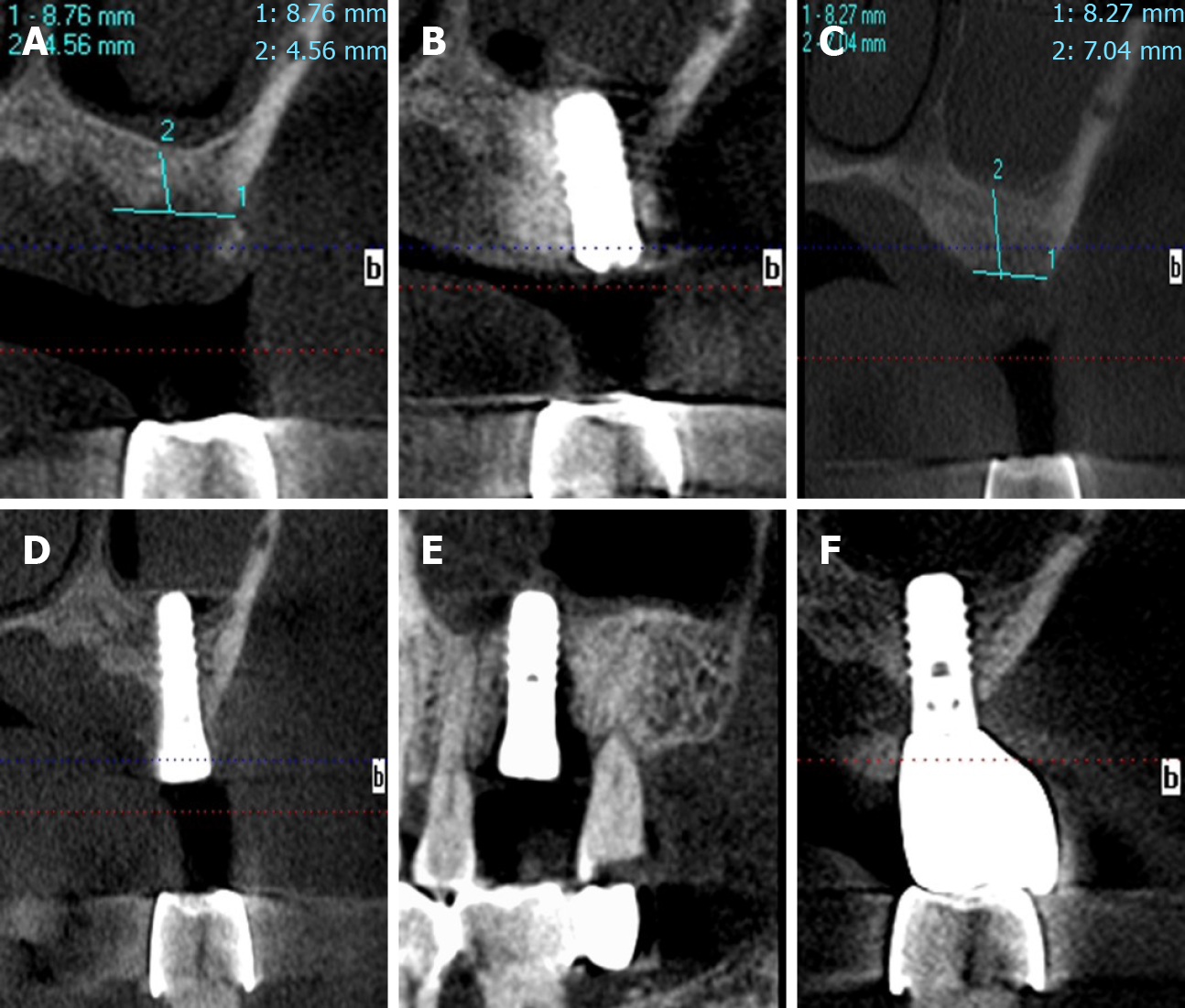
Figure 4 X-ray images of case 4.
A: In the preoperative X-ray image of the first-stage surgery, the residual bone height was 4.56 mm; B: In the postoperative X-ray image of the first-stage surgery, we placed a 4.8 mm-wide and 10 mm-long implant (Straumann); C: In the preoperative X-ray image of the second-stage surgery, the residual bone height was 7.04 mm; D: In the postoperative X-ray image of the second-stage surgery, we placed 4.8 mm-wide and 10 mm-long implant (Straumann); E: The X-ray image after prosthetic rehabilitation; F: The x-ray image after prosthetic rehabilitation.
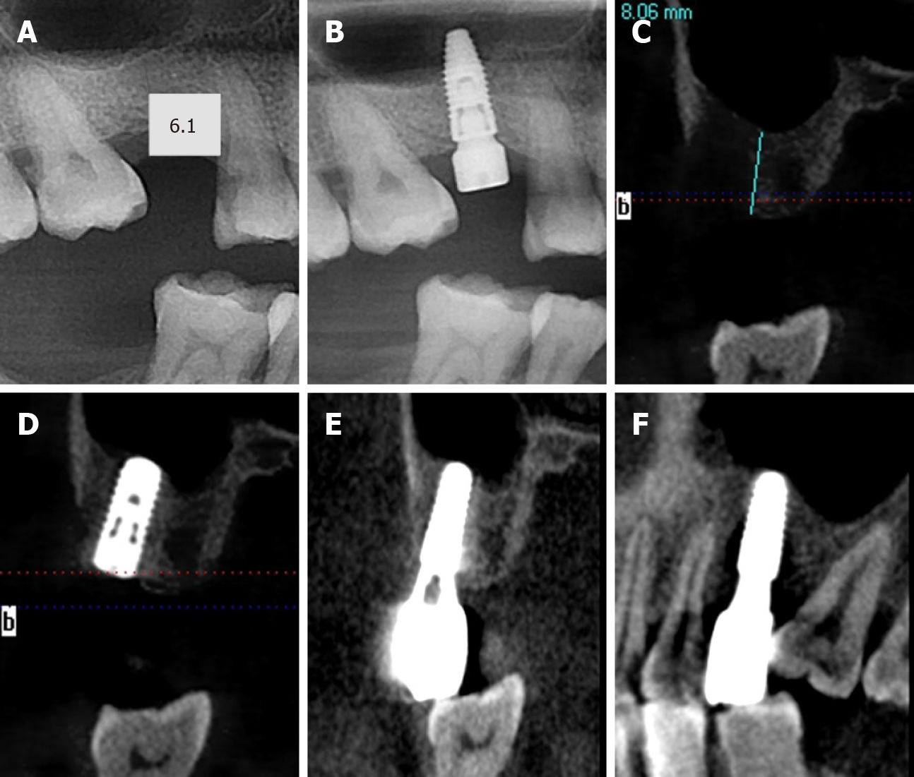
Figure 5 X-ray images of case 5.
A: In the preoperative X-ray image of the first-stage surgery, the residual bone height was 6.10 mm; B: In the postoperative X-ray image of the first-stage surgery, we placed a 4.3 mm-wide and 10 mm-long implant (Nobel); C: In the preoperative X-ray image of the second-stage surgery, the residual bone height was 8.06 mm; D: In the postoperative X-ray image of the second-stage surgery, we placed a 4.1 mm-wide and 10 mm-long implant (Straumann); E: The X-ray image after prosthetic rehabilitation; F: The X-ray image after prosthetic rehabilitation.
- Citation: Lin ZZ, Xu DQ, Ye ZY, Wang GG, Ding X. Two-stage transcrestal sinus floor elevation-insight into replantation: Six case reports. World J Clin Cases 2021; 9(10): 2386-2393
- URL: https://www.wjgnet.com/2307-8960/full/v9/i10/2386.htm
- DOI: https://dx.doi.org/10.12998/wjcc.v9.i10.2386


