Copyright
©The Author(s) 2021.
World J Clin Cases. Apr 6, 2021; 9(10): 2334-2343
Published online Apr 6, 2021. doi: 10.12998/wjcc.v9.i10.2334
Published online Apr 6, 2021. doi: 10.12998/wjcc.v9.i10.2334
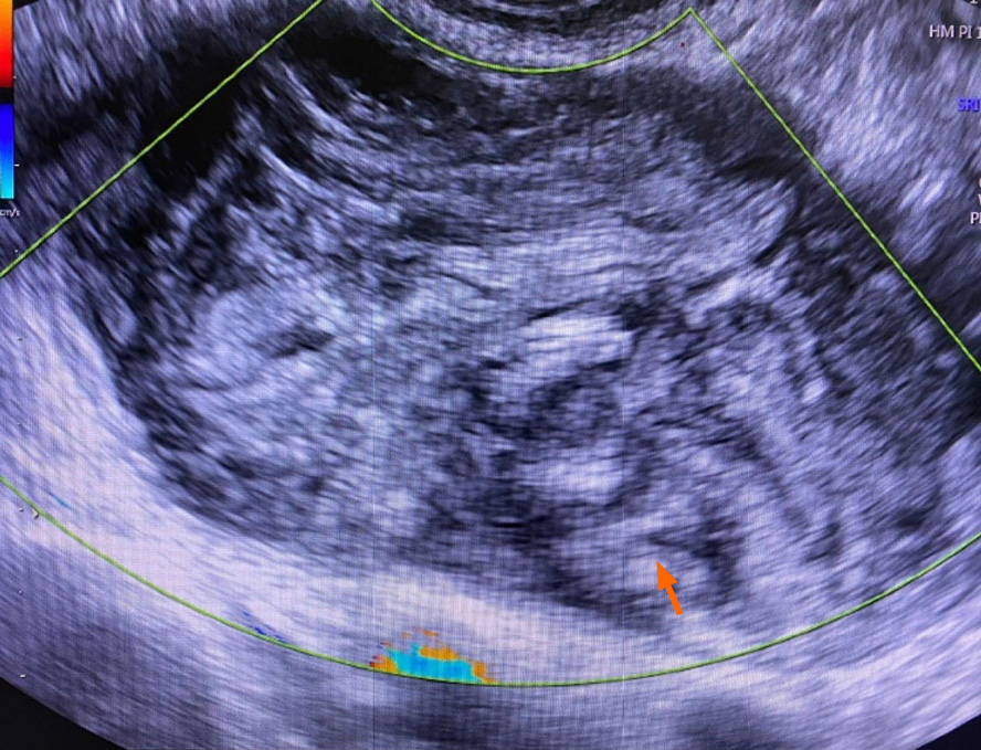
Figure 1
Low-grade mucinous appendiceal neoplasm presenting as a right adnexal mass (transvaginal ultrasound).
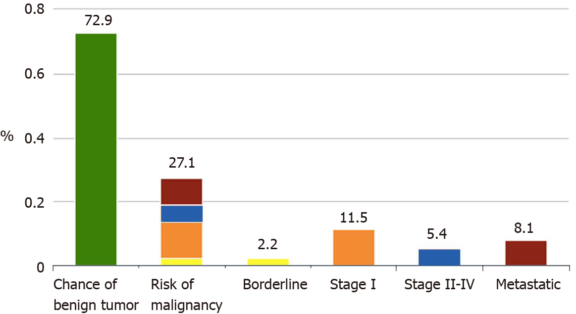
Figure 2
Low-grade mucinous appendiceal neoplasm assumed as an adnexal neoplasm: Results of the tumor assessment using the international ovarian tumor analysis ADNEX prediction model.
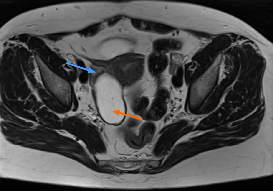
Figure 3 Low-grade mucinous appendiceal neoplasm mimicking an ovarian tumor (magnetic resonance imaging presentation).
The blue arrow indicates the right ovary; the orange arrow indicates the tumor apparently originating from the right ovary.
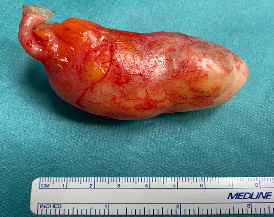
Figure 4
Low-grade mucinous appendiceal neoplasm macroscopic features (appendectomy with tumorectomy specimen).
Figure 5 Appendectomy with tumorectomy specimens (histological characteristics).
A: Proximal appendiceal stump with normal histological features [Hematoxylin & eosin (H&E) staining, 20 × magnitude]; B: Low-grade mucinous appendiceal neoplasm (LAMN) (H&E staining, 40 × magnitude, blue arrow showing acellular mucin); C: LAMN (H&E staining, 100 × magnitude, orange arrow absence of high-grade epithelial dysplasia).
- Citation: Borges AL, Reis-de-Carvalho C, Chorão M, Pereira H, Djokovic D. Low-grade mucinous appendiceal neoplasm mimicking an ovarian lesion: A case report and review of literature. World J Clin Cases 2021; 9(10): 2334-2343
- URL: https://www.wjgnet.com/2307-8960/full/v9/i10/2334.htm
- DOI: https://dx.doi.org/10.12998/wjcc.v9.i10.2334


