Copyright
©The Author(s) 2021.
World J Clin Cases. Apr 6, 2021; 9(10): 2302-2311
Published online Apr 6, 2021. doi: 10.12998/wjcc.v9.i10.2302
Published online Apr 6, 2021. doi: 10.12998/wjcc.v9.i10.2302
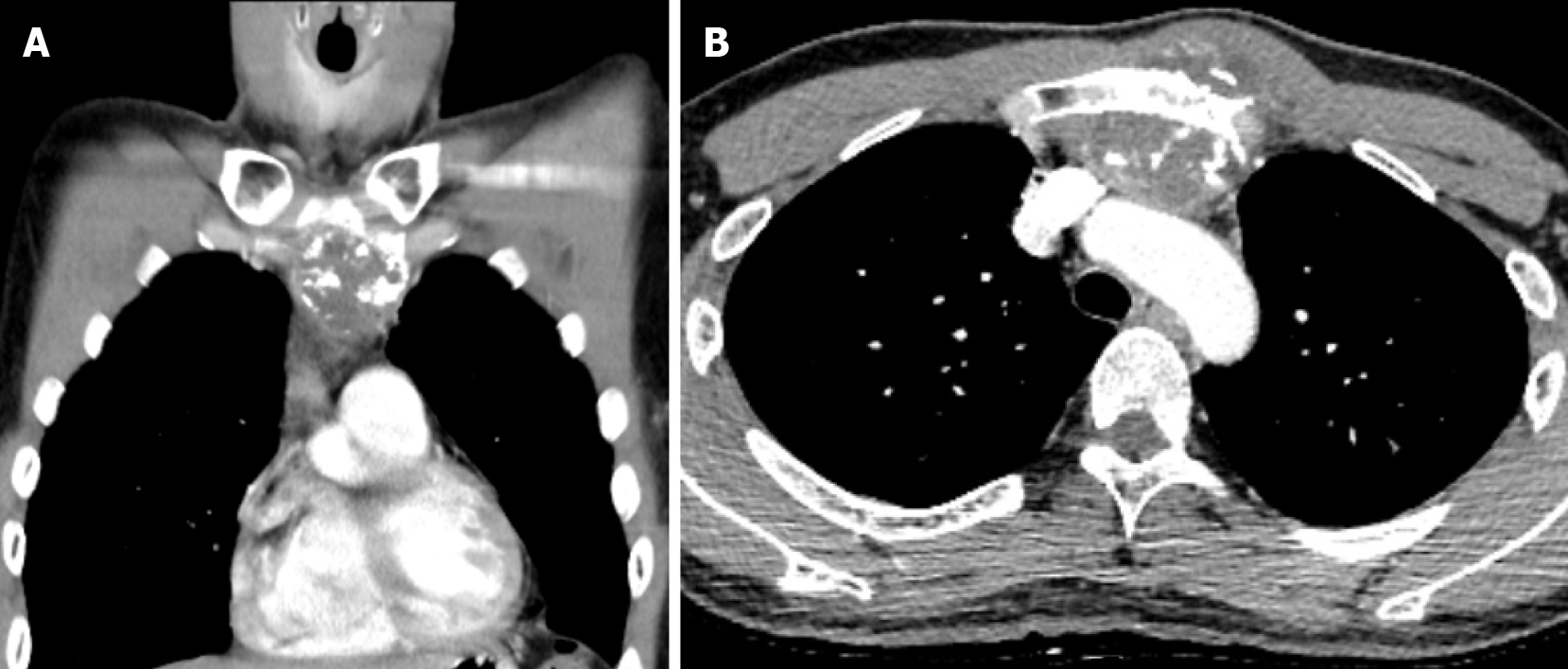
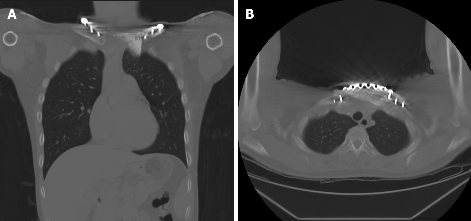
Figure 1 Focal osteoblastic mass in the sternum with bony exostosis and adjacent soft tissue calcification.
A and B: Coronal (A) and axial (B) views of contrast computed tomography.
Figure 2 Hypermetabolic mass overlying the upper sternum.
A and B: Three-dimensional reconstruction and axial view of positron emission tomography-computed tomography image.
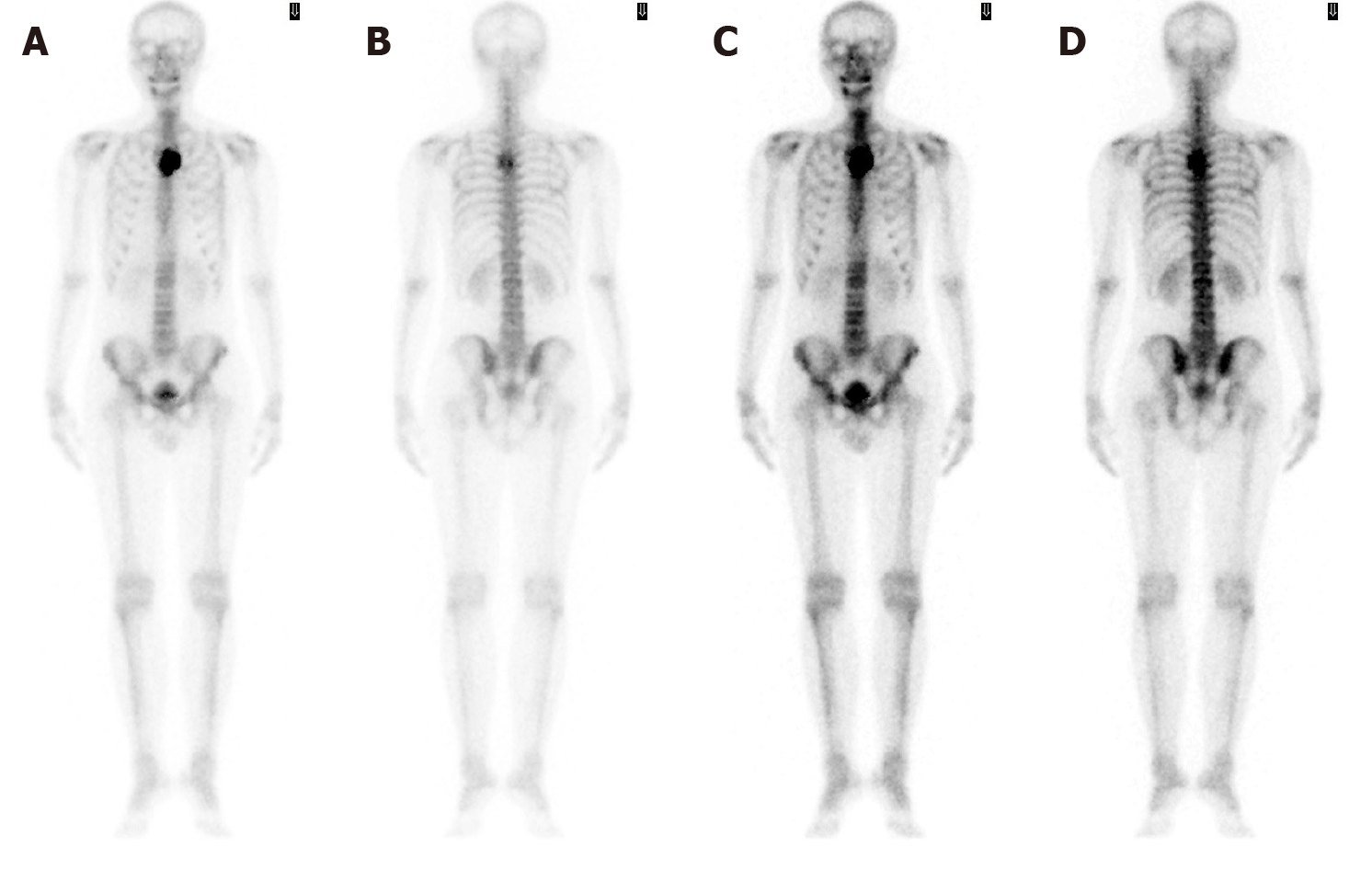
Figure 3 Bone scintigraphy revealing increased focal uptake in the sternum, showing a strong suspicion of a primary sternal tumor.
Figure 4 Focal ill-defined bony mass of the sternum with cortical destruction and periosteal reaction.
A-C: Magnetic resonance imaging with (A) coronal T2-weighted scan, (B) sagittal fat-suppressed T2-weighted scan, and (C) axial fat-suppressed T2-weighted scan.
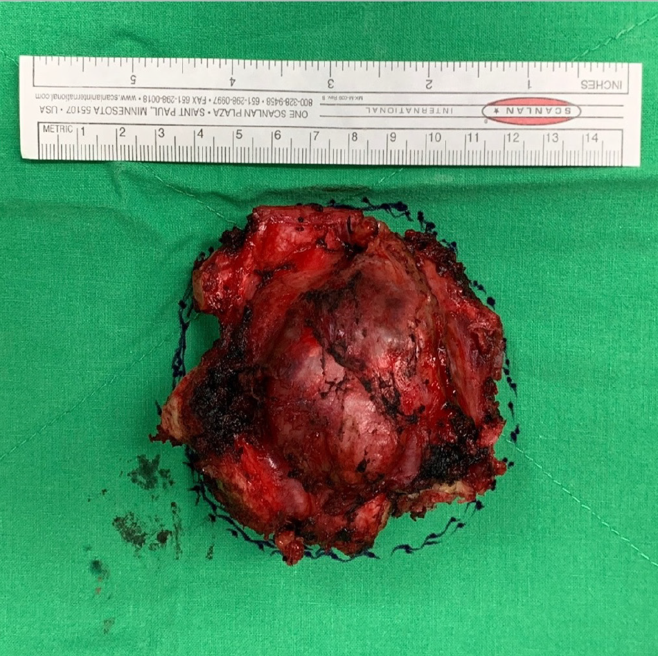
Figure 5 Gross specimen of the resected tumor showing a 6.
5 cm × 6.0 cm × 5.0 cm mass with a clean resected margin as confirmed by frozen section. The tumor comprised hyaline cartilage.
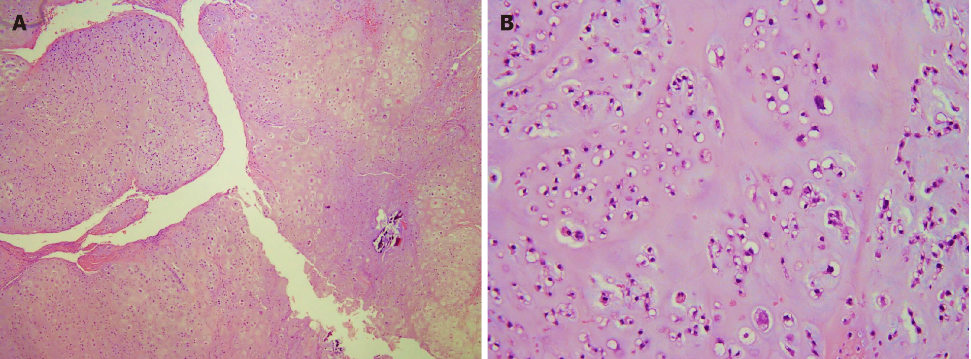
Figure 6 Pathological images.
A and B: Low power (A, 40 ×) and high power (B, 200 ×) histopathology pictures of chondrosarcoma showing hyaline cartilage matrix and chondrocytes in lacunae.
Figure 7 Intraoperative photos demonstrating the shaping of bone cement spacer for the sternum.
A: We measured the defect size carefully, remodeled the polymethyl methacrylate to meet the shape of manubrium, and inserted Kirschner wire creating holes for anchoring sutures; B: Mesh placement over the mediastinum to prevent cement spacer from direct contact with the surrounding musculature and major blood vessels; C: Ethibond sutures passing through the spacer anchoring surrounding bony structures, muscles, and the J-shaped locking plate.
Figure 8 Coronal and axial views of computed tomography at 1-year follow-up revealing absence of local tumor recurrence.
A: Coronal; B: Axial.
- Citation: Lin CW, Ho TY, Yeh CW, Chen HT, Chiang IP, Fong YC. Innovative chest wall reconstruction with a locking plate and cement spacer after radical resection of chondrosarcoma in the sternum: A case report. World J Clin Cases 2021; 9(10): 2302-2311
- URL: https://www.wjgnet.com/2307-8960/full/v9/i10/2302.htm
- DOI: https://dx.doi.org/10.12998/wjcc.v9.i10.2302


