Copyright
©The Author(s) 2021.
World J Clin Cases. Apr 6, 2021; 9(10): 2281-2288
Published online Apr 6, 2021. doi: 10.12998/wjcc.v9.i10.2281
Published online Apr 6, 2021. doi: 10.12998/wjcc.v9.i10.2281
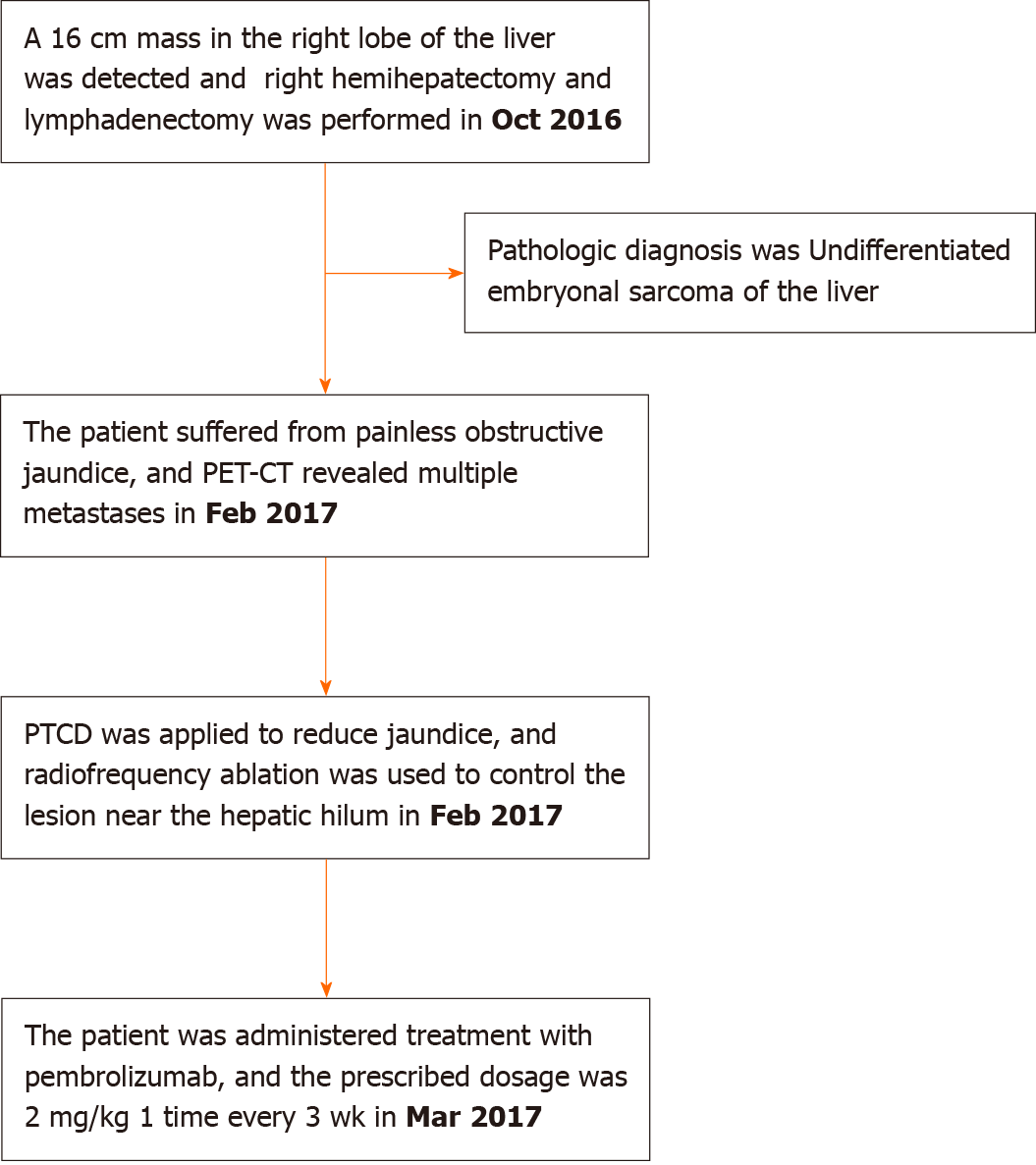
Figure 1 Timeline of the case.
PET-CT: Positron emission tomography-computed tomography; PTCD: Percutaneous transhepatic cholangial drainage.
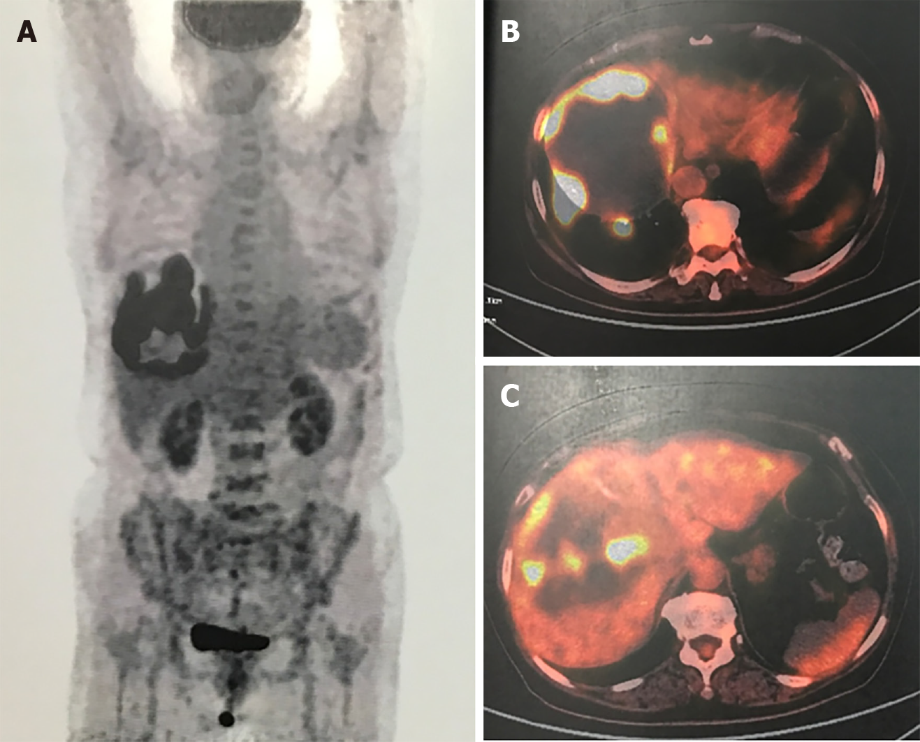
Figure 2 Positron emission tomography-computed tomography.
A-C: Radioactive concentration of 18F-fluorodeoxyglucose on the tumor rim, and no radioactive concentration was observed in other areas of the body.
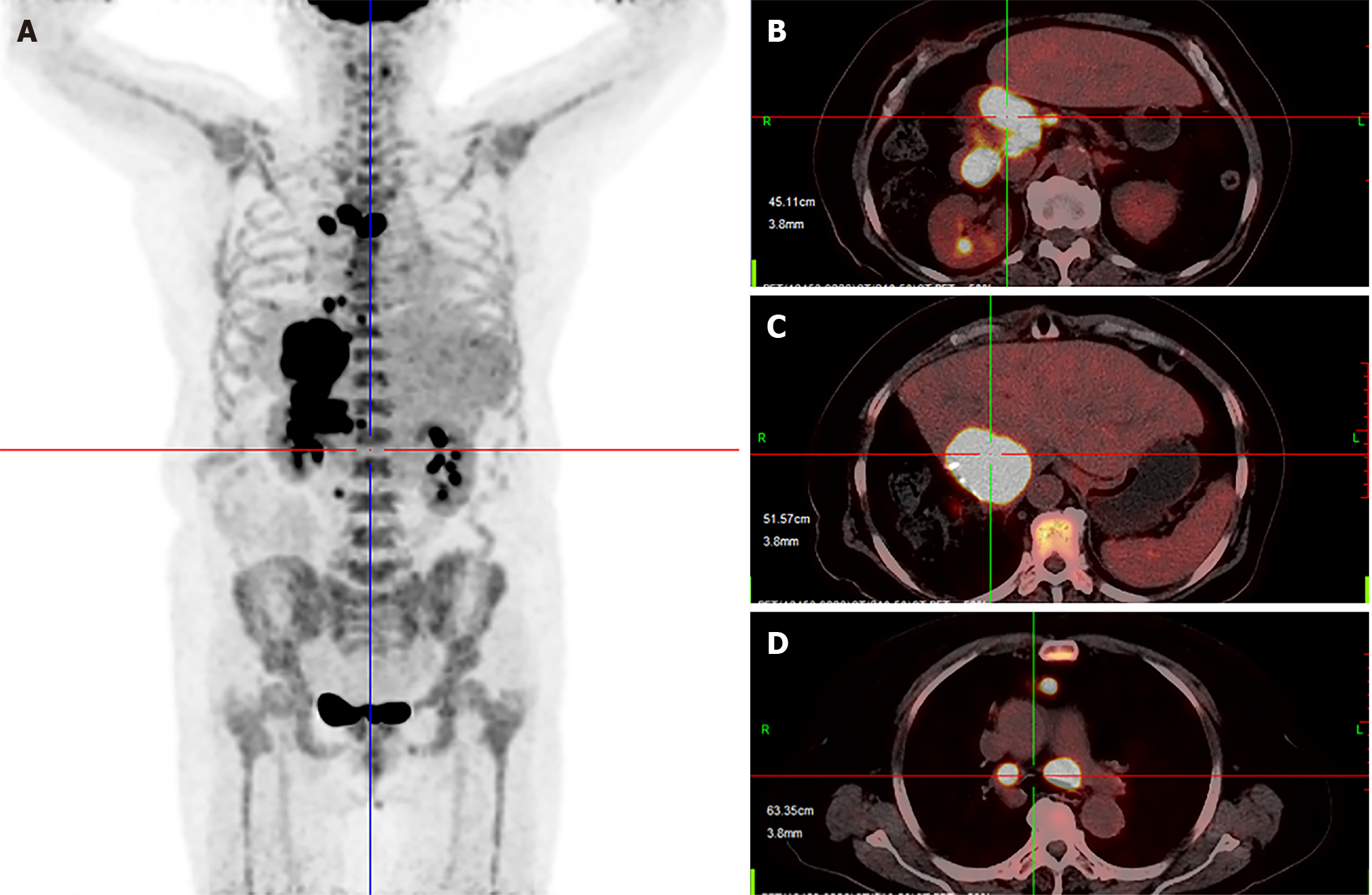
Figure 3 Positron emission tomography-computed tomography.
A-D: Multiple metastases located in the liver, mediastinum and peritoneum.
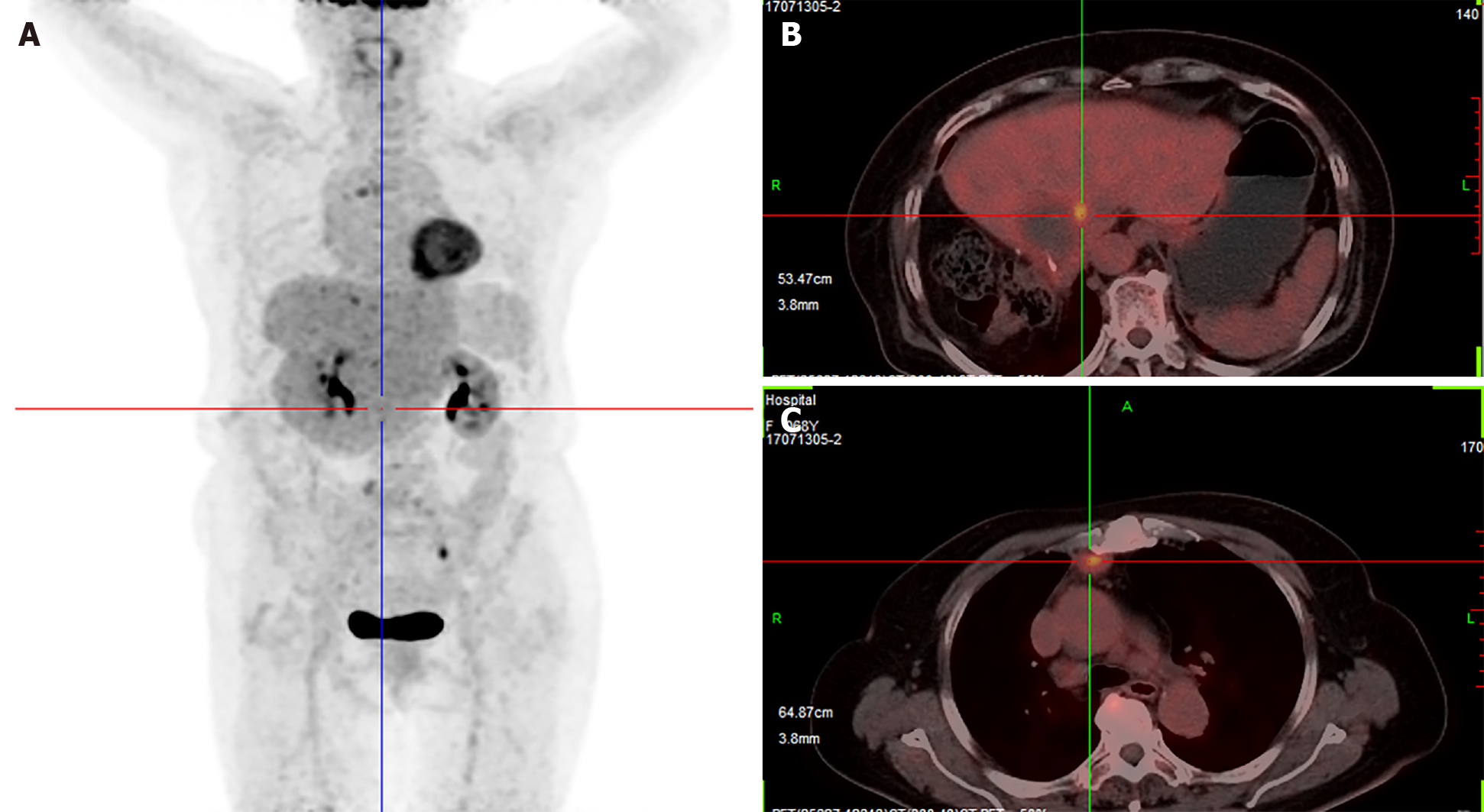
Figure 4 Positron emission tomography-computed tomography.
A-C: Multiple metastases had nearly disappeared.
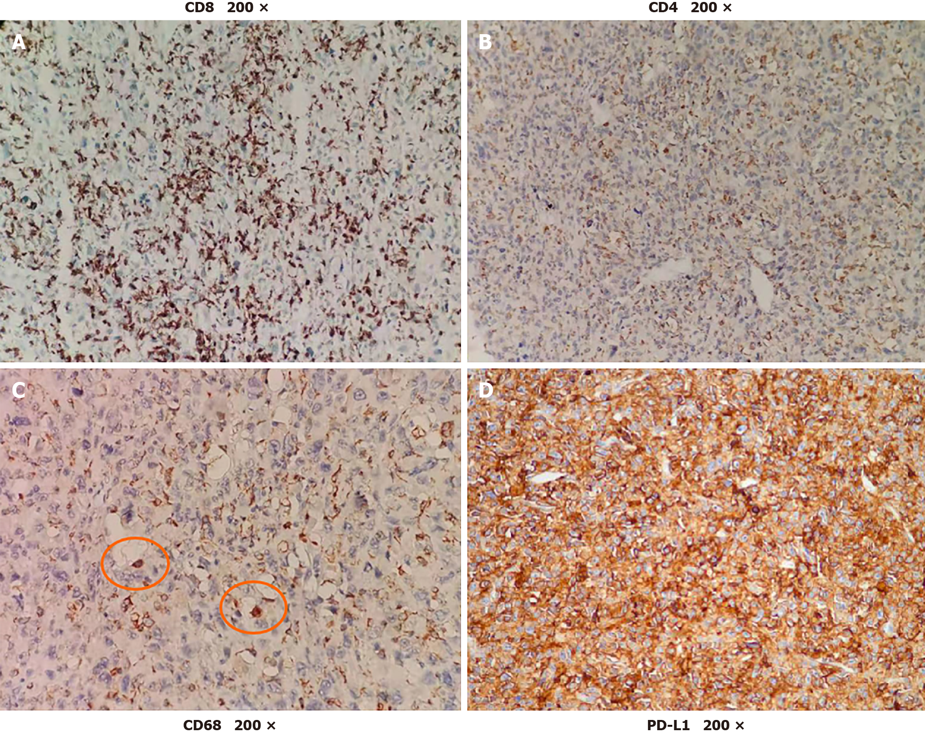
Figure 5 Immunohistological staining.
A-D: There was high expression of CD8, low expression of CD4 and little expression of CD68 in the tumor. Up to 90% of tumor cells expressed programmed death ligand 1 (PD-L1).
- Citation: Yu XH, Huang J, Ge NJ, Yang YF, Zhao JY. Recurrent undifferentiated embryonal sarcoma of the liver in adult patient treated by pembrolizumab: A case report. World J Clin Cases 2021; 9(10): 2281-2288
- URL: https://www.wjgnet.com/2307-8960/full/v9/i10/2281.htm
- DOI: https://dx.doi.org/10.12998/wjcc.v9.i10.2281


