Copyright
©The Author(s) 2021.
World J Clin Cases. Apr 6, 2021; 9(10): 2274-2280
Published online Apr 6, 2021. doi: 10.12998/wjcc.v9.i10.2274
Published online Apr 6, 2021. doi: 10.12998/wjcc.v9.i10.2274
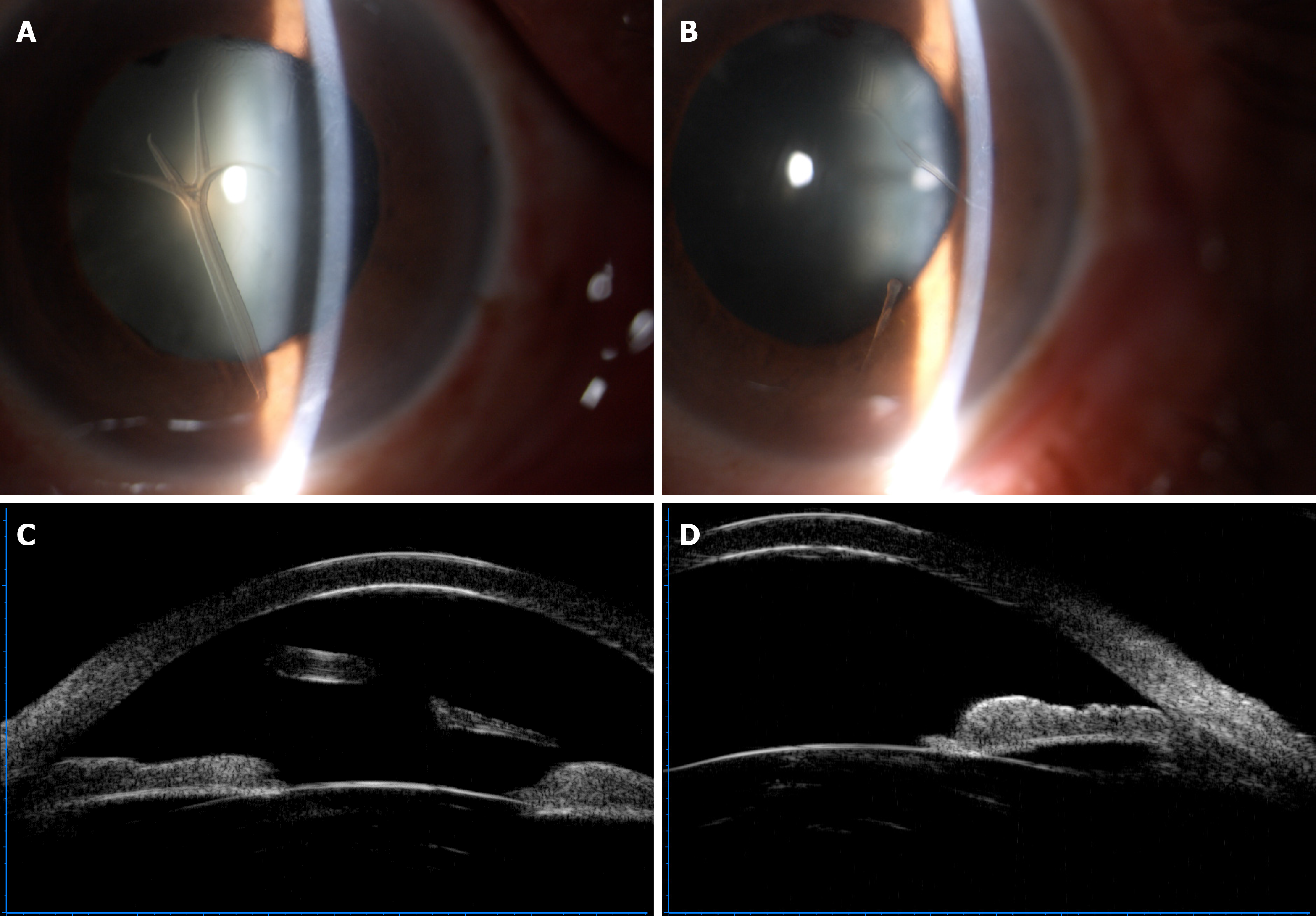
Figure 1 Appearance of retrocorneal scrolls under slit-lamp microscopy and ultrasonic biological microscopy.
A-D: An antler shaped scroll extended to the anterior chamber and attached to the posterior surface of the cornea by a stalk in the right eye (A), and rod-like scrolls adhered to the corneal endothelium in the left eye (B). Free end of the scroll in the left eye (C) and adhered scroll in the right eye (D) with increased reflectivity of the posterior surface of the corneas were observed by ultrasonic biological microscopy.
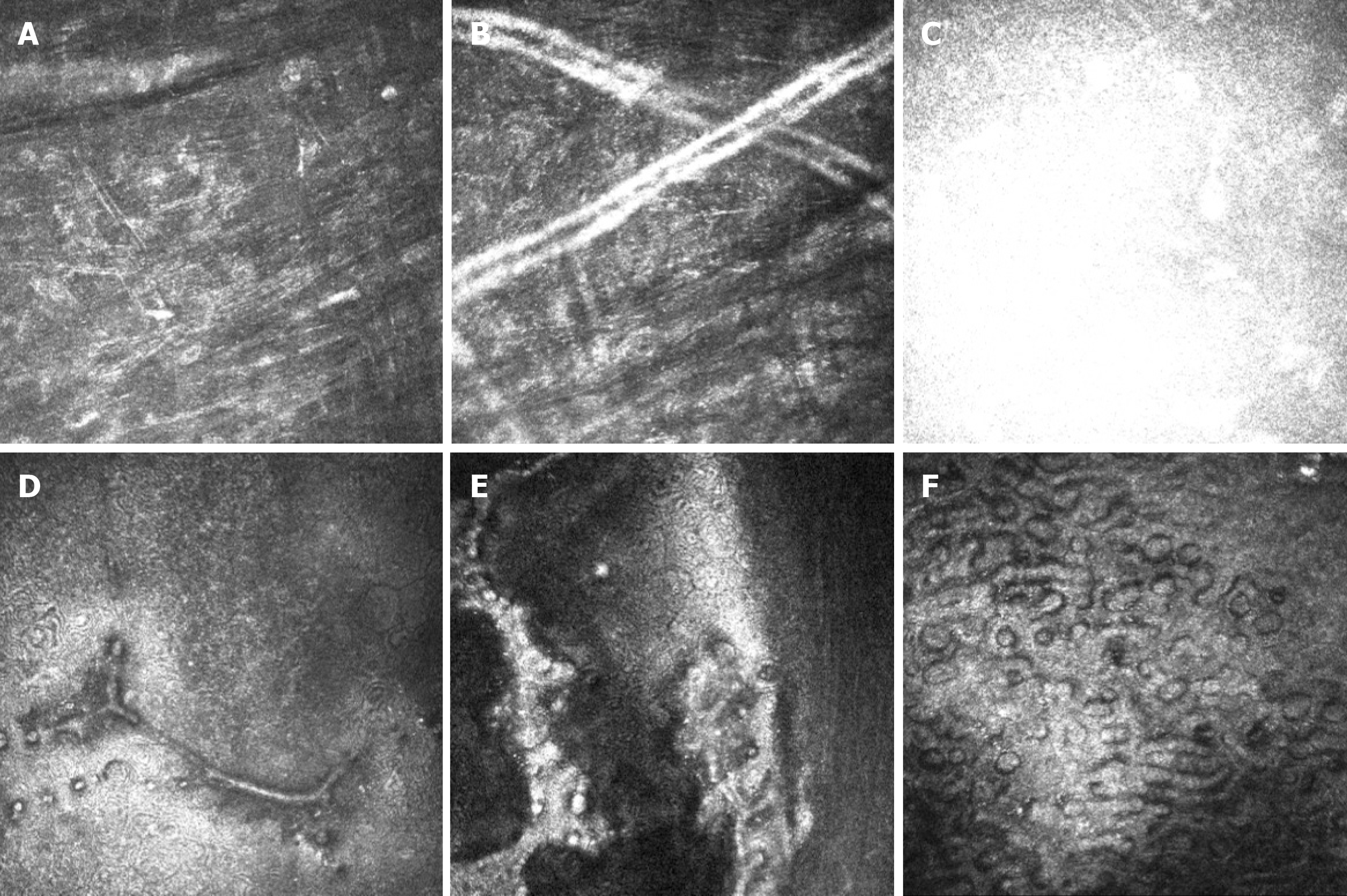
Figure 2 In vivo confocal microscopy showing the changes in the corneas.
A: Activated keratocytes and the altered structure of the extracellular tissue, manifested as thin bright lines in the stroma; B: Tubular structures in the posterior stroma; C: Highly reflective acellular structure at the level of the Descemet’s membrane; D and E: Strip confluent guttae at the level of the corneal endothelium and sheet-like confluent guttae adhesion to the endothelium on the left eye; F: Numerous confluent guttae and enlarged corneal endothelium on the right eye.
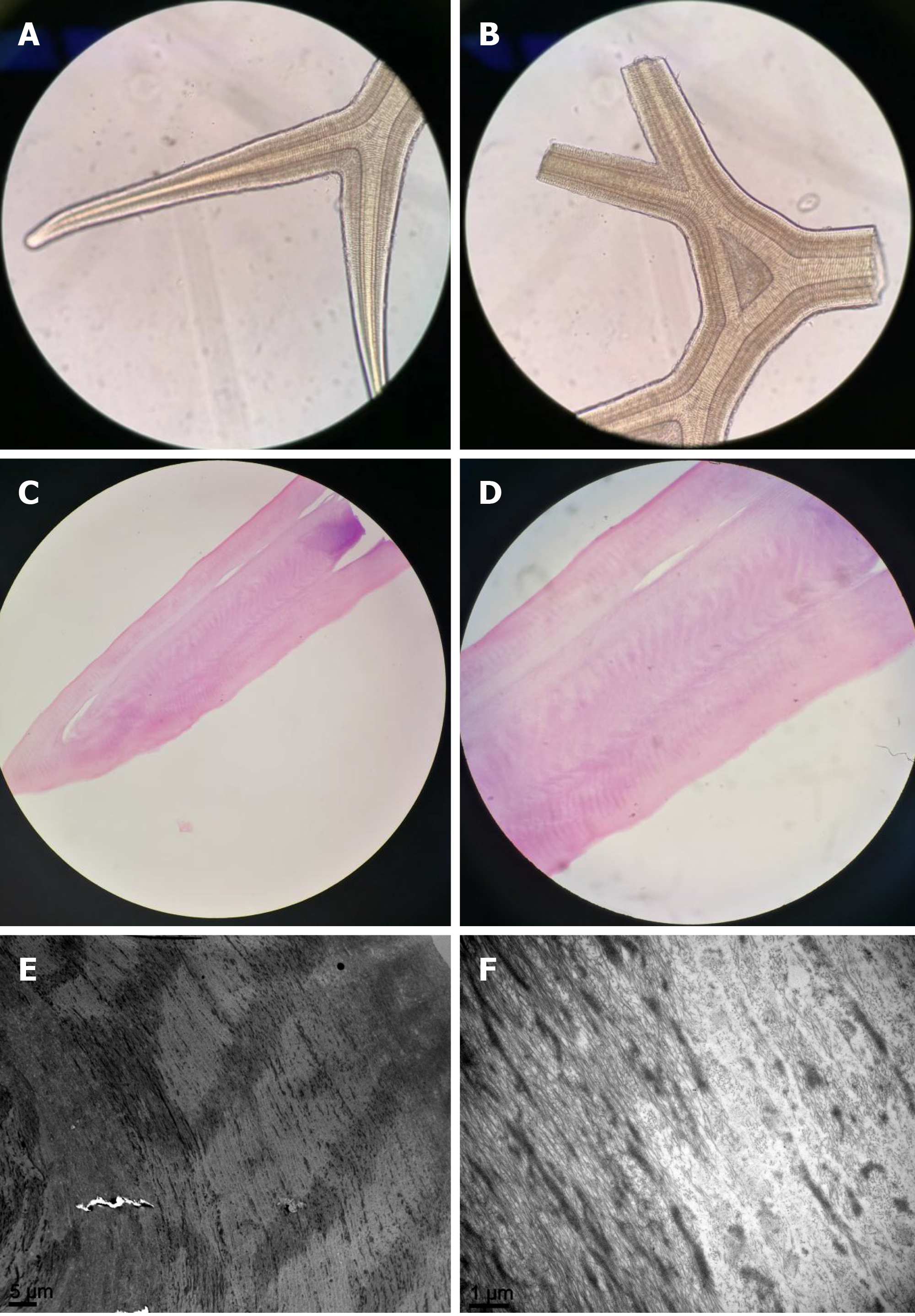
Figure 3 Gross and histologic images.
A and B: Gross microscopic examination showing the scrolls as regular acellular structure like antlers (× 40); C and D: Hematoxylin-eosin staining demonstrated that the scroll comprised eosinophilic acellular tissue (C, × 40; D, × 100); E and F: Scanning electron microscopy showed that the scroll was composed of similar fibrous tissue with a circular arrangement (E, × 2000; F, × 15000).
- Citation: Jin YQ, Hu YP, Dai Q, Wu SQ. Bilateral retrocorneal hyaline scrolls secondary to asymptomatic congenital syphilis: A case report. World J Clin Cases 2021; 9(10): 2274-2280
- URL: https://www.wjgnet.com/2307-8960/full/v9/i10/2274.htm
- DOI: https://dx.doi.org/10.12998/wjcc.v9.i10.2274


