Copyright
©The Author(s) 2021.
World J Clin Cases. Apr 6, 2021; 9(10): 2218-2227
Published online Apr 6, 2021. doi: 10.12998/wjcc.v9.i10.2218
Published online Apr 6, 2021. doi: 10.12998/wjcc.v9.i10.2218
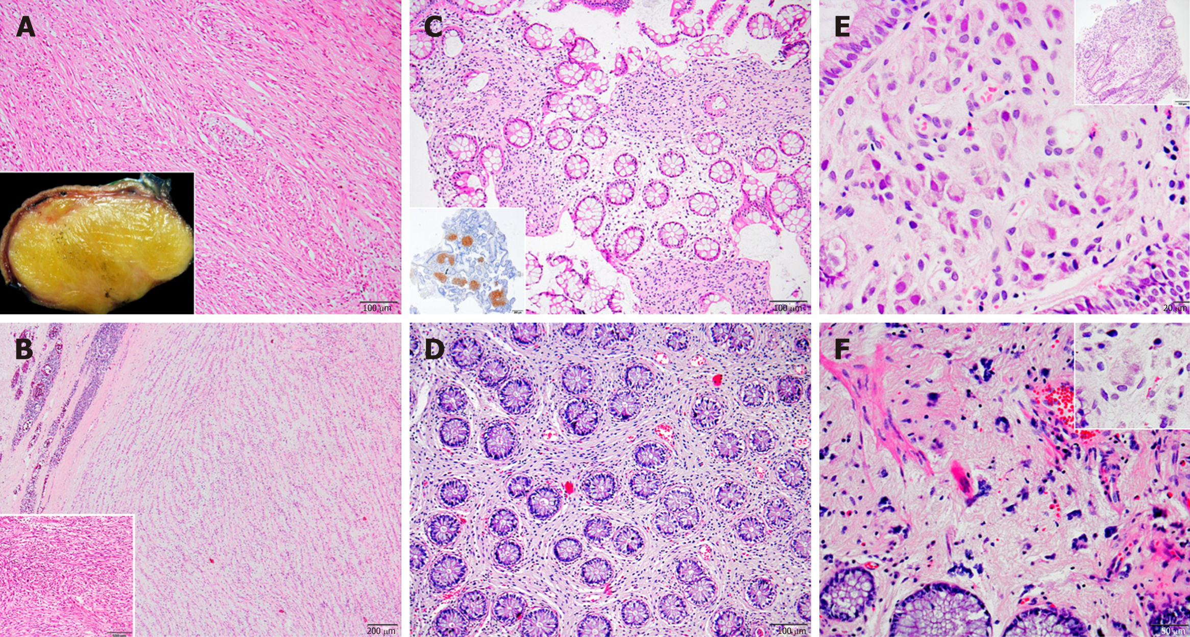
Figure 1 Histopathology of schwannomas, neurofibromatosis and ganglioneuromas.
A: Low power microscopic view of schwannoma [hematoxylin and eosin (H&E), × 100]; inset: Macroscopic appearance of a well circumscribed yellow-colored mass with an homogeneous cut surface; B: Schwannoma with Antoni A and B areas, showing a well-demarcated tumor with surrounding lymphoid aggregates (H&E, × 40); inset: cellular schwannoma (H&E, × 100); C: Patchy mucosal involvement of colonic neurofibromatosis (H&E, × 100); inset: a patchy S100 positivity is seen in the lamina propria (immunohistochemistry, × 40); D: Diffuse involvement of colonic neurofibromatosis, with an expansion of lamina propria mimicking a fibrotic process (H&E, × 100); E: Ganglioneuroma with a cluster of ganglion cells (H&E, × 400); inset: Low power view of ganglioneuroma expanding the lamina propria (H&E, × 100); F: Ganglioneuroma with scattered mildly dysmorphic ganglion cells in a pale eosinophilic Schwannian stroma (H&E, × 200); inset: mature ganglion cells consisting of compact, eosinophilic cytoplasm with distinct cell borders and a single eccentric nucleus with a prominent nucleolus (H&E, × 400).
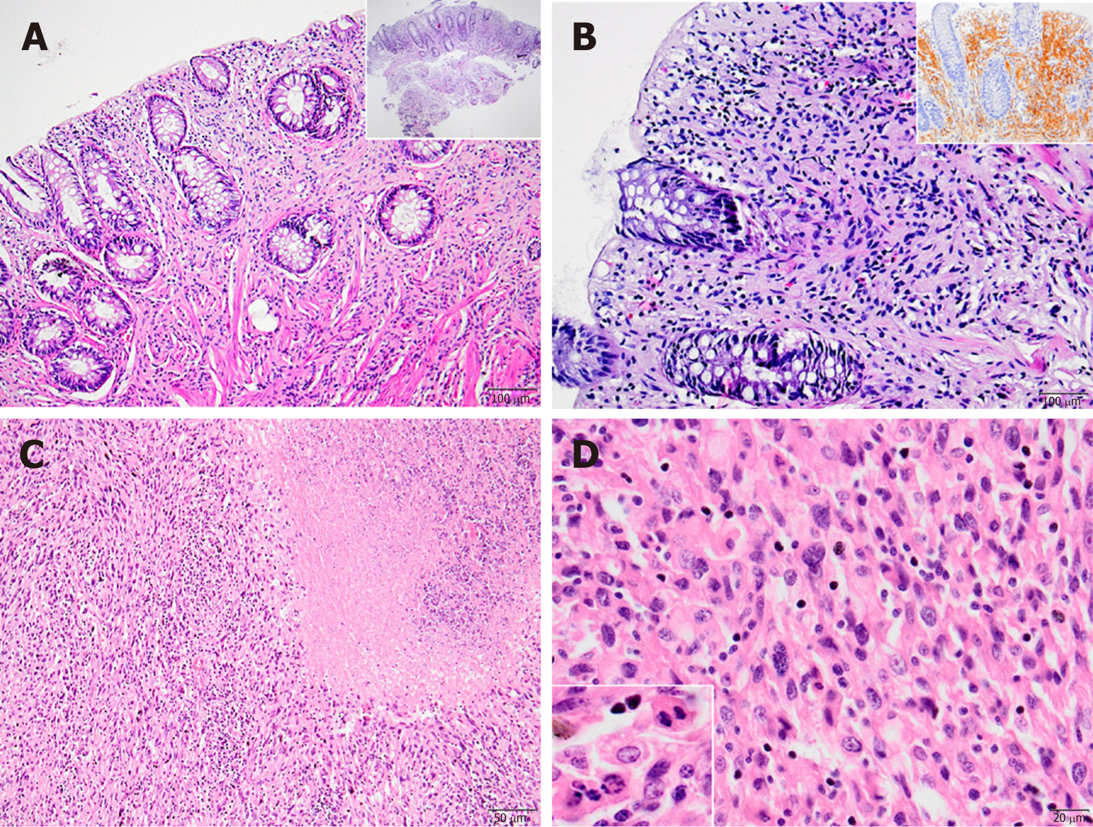
Figure 2 Histopathology of Schwann cell hamartoma and malignant peripheral sheet tumor.
A: Low (inset) and high power view of mucosal Schwann cell hamartoma located in sigmoid colon [hematoxylin and eosin (H&E), × 40 and × 100, respectively]; B: Mucosal Schwann cell hamartoma: Benign mucosal proliferation of Schwann cells filling and expanding the lamina propria with poorly circumscribed margins (H&E, × 200); inset: diffuse S100 positivity supporting the Schwannian origin (immunohistochemistry, × 100); C: Malignant peripheral nerve sheet tumor with broad geographic necrosis (H&E, × 100); D: Prominent cellular pleomorphism (H&E, × 400) and frequent mitosis and apoptotic bodies seen in malignant peripheral nerve sheet tumor (inset: H&E, × 400).
- Citation: Simsek C, Uner M, Ozkara F, Akman O, Akyol A, Kav T, Sokmensuer C, Gedikoglu G. Comprehensive clinicopathologic characteristics of intraabdominal neurogenic tumors: Single institution experience. World J Clin Cases 2021; 9(10): 2218-2227
- URL: https://www.wjgnet.com/2307-8960/full/v9/i10/2218.htm
- DOI: https://dx.doi.org/10.12998/wjcc.v9.i10.2218


