Copyright
©The Author(s) 2021.
World J Clin Cases. Jan 6, 2021; 9(1): 284-290
Published online Jan 6, 2021. doi: 10.12998/wjcc.v9.i1.284
Published online Jan 6, 2021. doi: 10.12998/wjcc.v9.i1.284
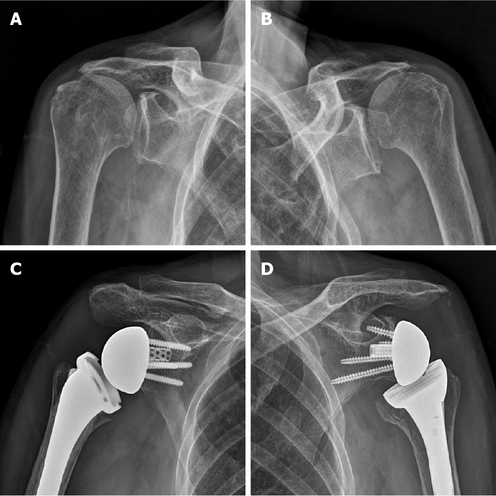
Figure 1 Preoperative (A and B) and postoperative (C and D) plain radiographs of both shoulder joints.
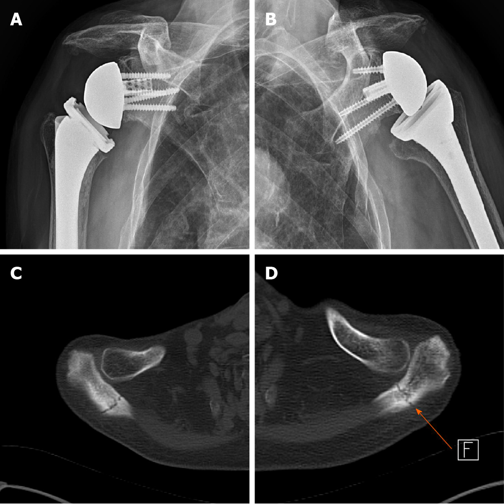
Figure 2 Plain radiographs and computed tomography scan of both shoulder joints show non-displaced acromial base fracture (Both Levy type II fracture).
A: Right shoulder plain radiograph; B: Left shoulder plain radiograph; C: Right shoulder computed tomography (CT) scan; D: Left shoulder CT scan.
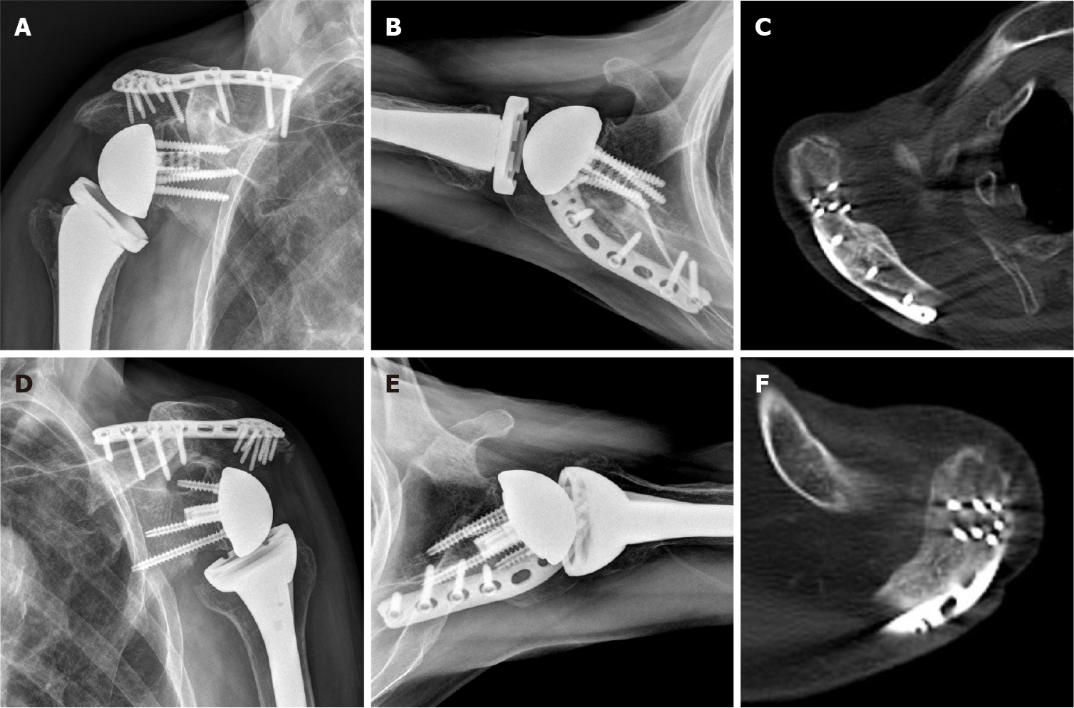
Figure 3 Plain radiographs and computed tomography scan at 2 years after plate fixation show complete healing of the fracture.
A and B: Right shoulder plain radiographs; C: Right shoulder computed tomography (CT) scan; D and E: Left shoulder plain radiographs; F: Left shoulder CT scan.
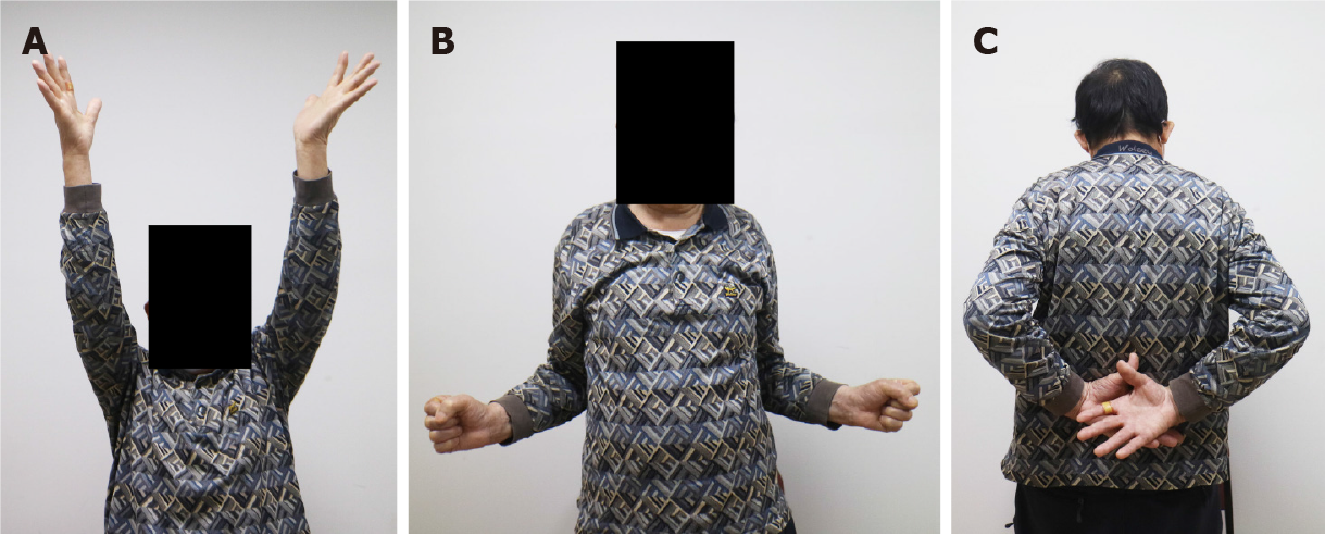
Figure 4 Clinical photos for active range of shoulder motion at 2 years after plate fixation (A-C).
- Citation: Kim DH, Kim BS, Cho CH. Simultaneous bilateral acromial base fractures after staged reverse total shoulder arthroplasty: A case report. World J Clin Cases 2021; 9(1): 284-290
- URL: https://www.wjgnet.com/2307-8960/full/v9/i1/284.htm
- DOI: https://dx.doi.org/10.12998/wjcc.v9.i1.284


