Copyright
©The Author(s) 2021.
World J Clin Cases. Jan 6, 2021; 9(1): 278-283
Published online Jan 6, 2021. doi: 10.12998/wjcc.v9.i1.278
Published online Jan 6, 2021. doi: 10.12998/wjcc.v9.i1.278
Figure 1 Tumor imaging and excision.
A: Contrast-enhanced computed tomography of the abdomen reveals an irregular mass at the left spermatic cord extending to the epididymis; B: Intraoperative photograph of the indurated mass measuring 4.0 cm × 1.7 cm on the spermatic cord and epididymis, without invasion to the testis. A biopsy was obtained from the spot indicated by the pointer.
Figure 2 Ipsilateral hydronephrosis.
A: Retrograde lymphatic metastasis-related left hydronephrosis in November 2019; B: Left hydronephrosis showed regression in March 2020, after systemic chemotherapy.
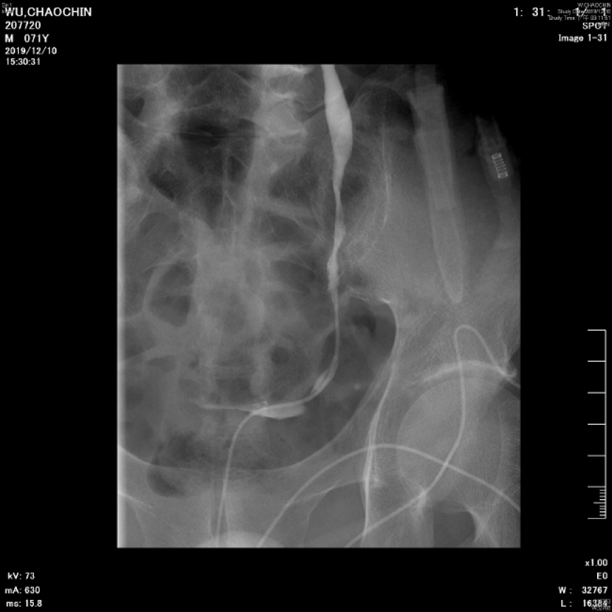
Figure 3 Left ureteral stenosis.
Left retrograde pyelography showed multiple stenosis and narrowing points along middle to lower ureter, which led to left hydronephrosis and hydroureter.
Figure 4 Histopathology.
A: Hematoxylin and eosin–stained section showed fibrovascular tissue with infiltration of nests of poorly differentiated adenocarcinoma; B: The immunohistochemically stained section was diffusely and strongly positive for CDX2, which is a marker indicative of adenocarcinoma of intestinal origin.
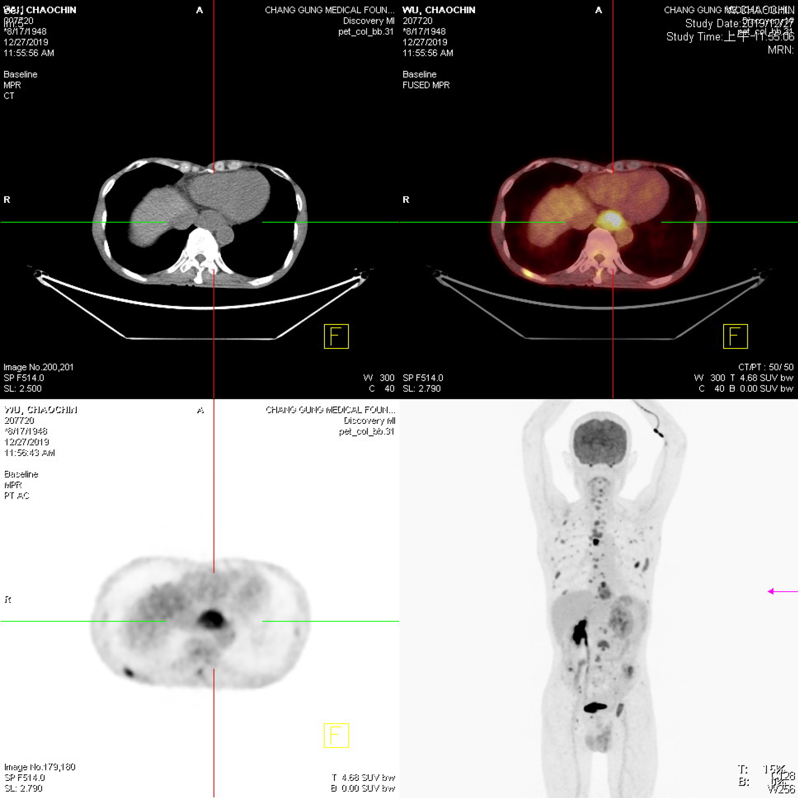
Figure 5 Tumor scan.
Whole-body tumor scan from the skull vertex to upper thighs was performed at 60 min after injection of 18F-fluorodeoxyglucose. Purple arrow indicates gastric stump recurrence. Multiple faint nodules were observed in the pelvic cavity, suggesting peritoneal dissemination, as well as the cause of ureteral stenosis.
- Citation: Tsao SH, Chuang CK. Krukenberg tumor with concomitant ipsilateral hydronephrosis and spermatic cord metastasis in a man: A case report. World J Clin Cases 2021; 9(1): 278-283
- URL: https://www.wjgnet.com/2307-8960/full/v9/i1/278.htm
- DOI: https://dx.doi.org/10.12998/wjcc.v9.i1.278


