Copyright
©The Author(s) 2021.
World J Clin Cases. Jan 6, 2021; 9(1): 245-251
Published online Jan 6, 2021. doi: 10.12998/wjcc.v9.i1.245
Published online Jan 6, 2021. doi: 10.12998/wjcc.v9.i1.245
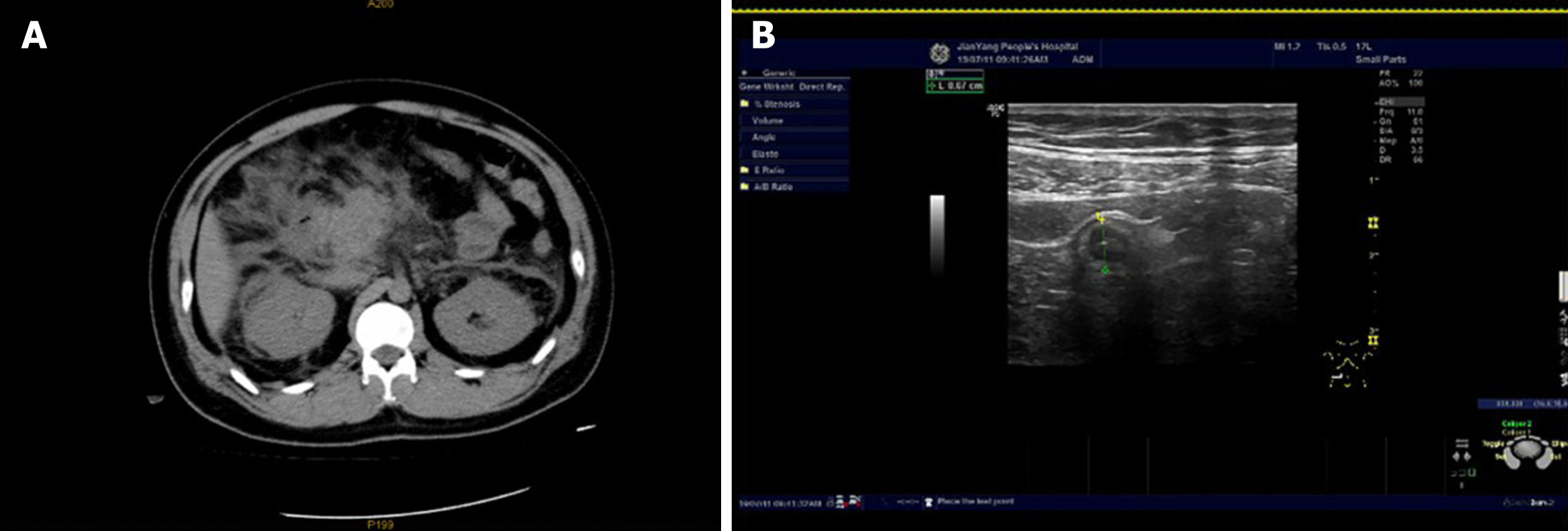
Figure 1 On-contrast computed tomography manifestations and Doppler ultrasound of Case 1.
A: On-contrast computed tomography manifestations showing features of acute pancreatitis with extensive peripancreatic fat stranding; B: Abdominal color Doppler ultrasound showing a dilated and thickened appendix.
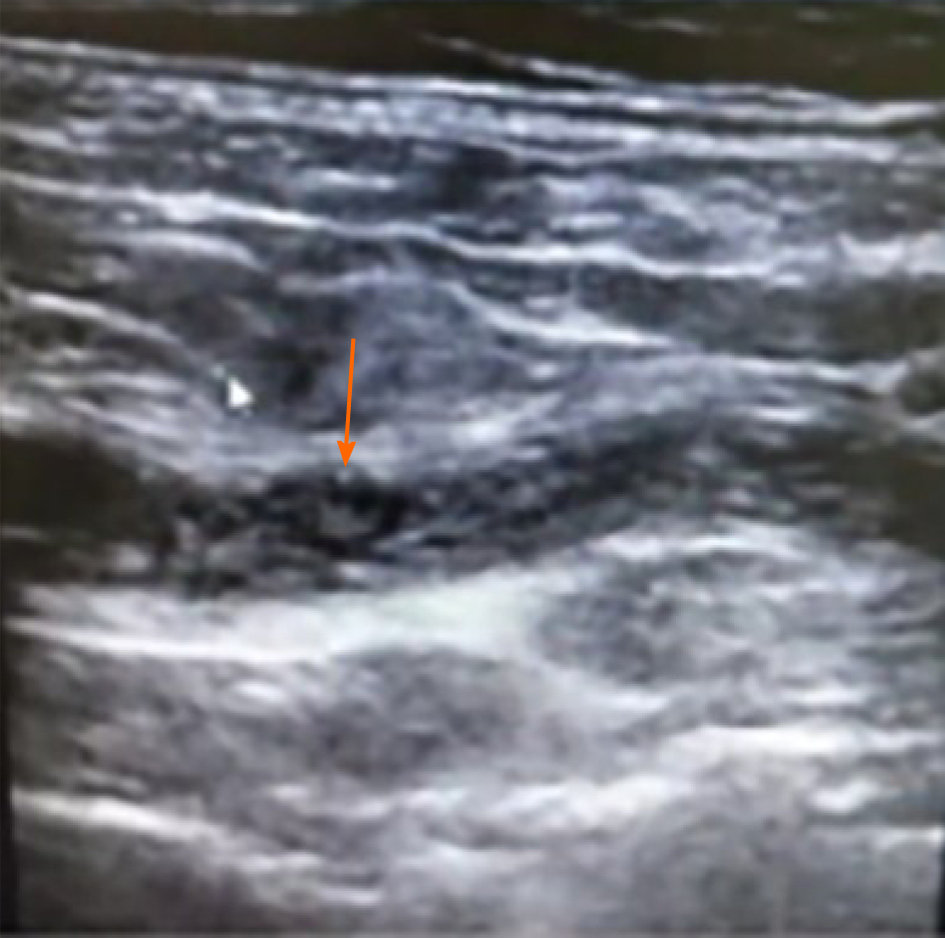
Figure 2 B-ultrasound of Case 2 showed the dilated appendiceal cavity with the presence of cord-like sediments inside.
Figure 3 Course of treatment in Case 1.
A: Conical transparent cap fixed to the appendiceal opening; B: Retrograde appendicography performed during endoscopic retrograde appendicitis treatment; C: Flushing of the appendix showed pus outflow.
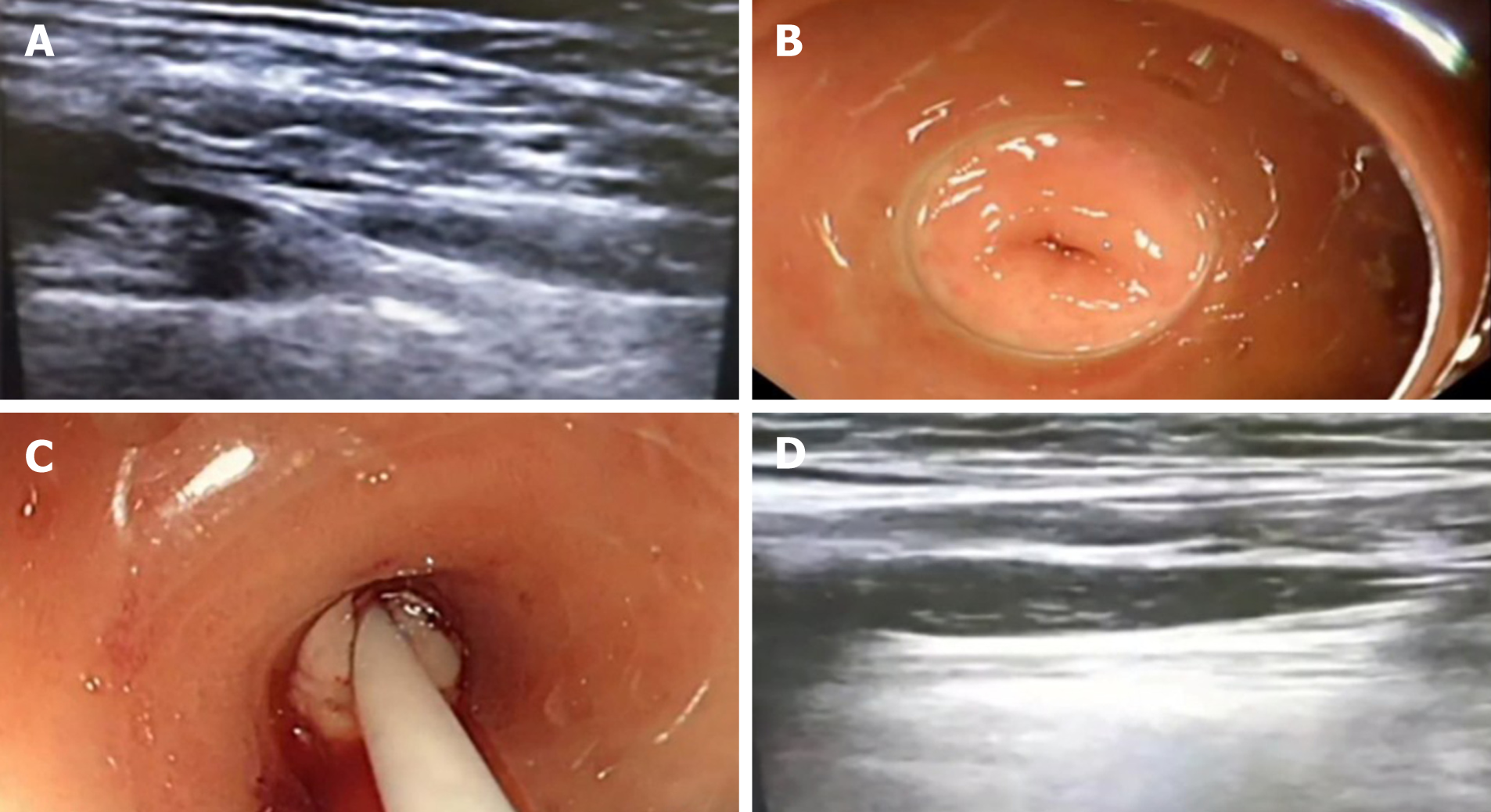
Figure 4 Course of treatment in Case 2.
A: B-ultrasound showing the intestinal lens-end of the tapered cap; B: The tapered transparent cap at the lens-end of the intestine enters the opening of the appendix; C: Flushing of the appendix showed pus outflow; D: Irrigation of the appendiceal cavity under ultrasound guidance.
- Citation: Du ZQ, Ding WJ, Wang F, Zhou XR, Chen TM. Endoscopic treatment for acute appendicitis with coexistent acute pancreatitis: Two case reports. World J Clin Cases 2021; 9(1): 245-251
- URL: https://www.wjgnet.com/2307-8960/full/v9/i1/245.htm
- DOI: https://dx.doi.org/10.12998/wjcc.v9.i1.245


