Copyright
©The Author(s) 2021.
World J Clin Cases. Jan 6, 2021; 9(1): 236-244
Published online Jan 6, 2021. doi: 10.12998/wjcc.v9.i1.236
Published online Jan 6, 2021. doi: 10.12998/wjcc.v9.i1.236
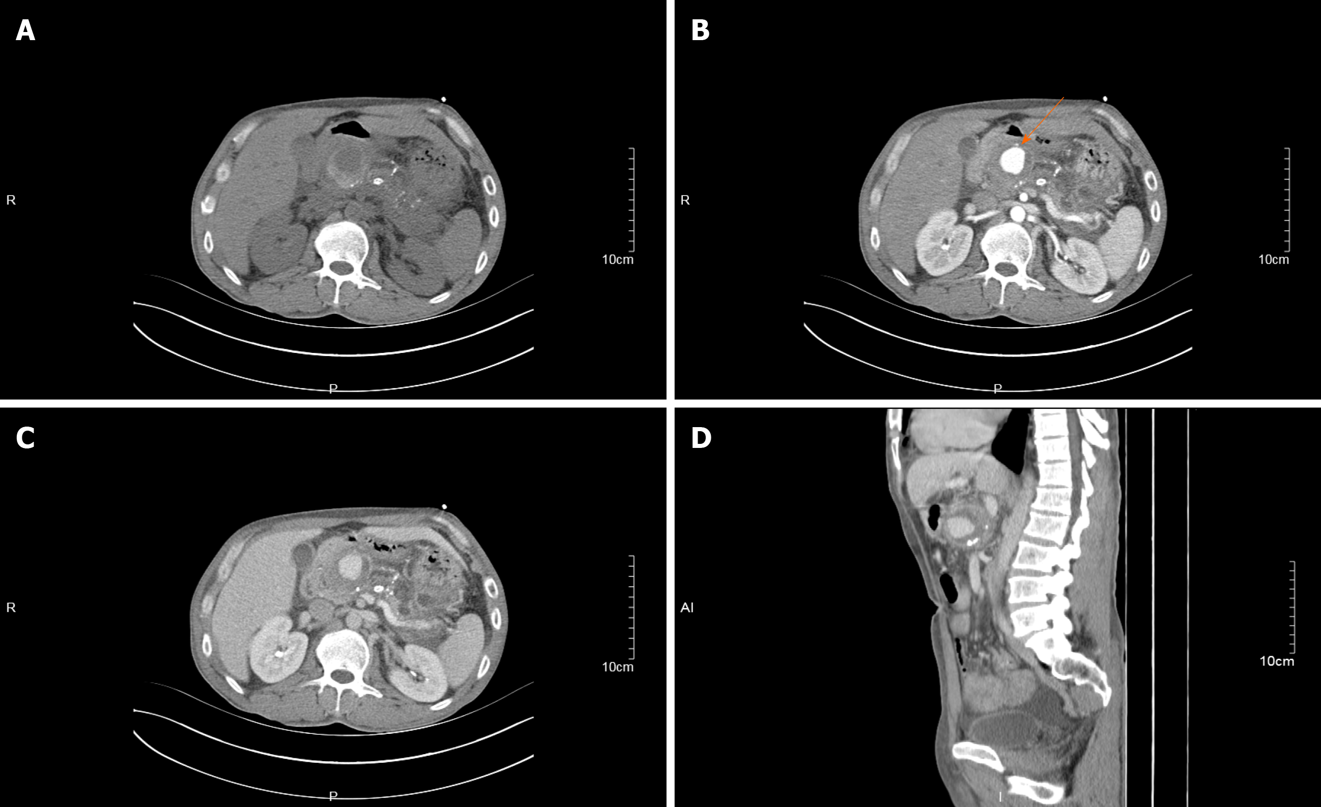
Figure 1 Contrast-enhanced computed tomography of the abdomen.
A: Noncontrast-enhanced computed tomography revealed pancreatic swelling, multiple granular calcifications in the parenchyma, pancreatic duct dilatation in the body and tail, and a circular hypointense opacity surrounded by a slightly hyperintense wall in the pancreatic head; B: Obvious enhancement in the pancreatic head lesion in the arterial phase, with a computed tomography value of approximately 195 HU and without enhancement of the surrounding tissue. The lesion is closely related to the gastroduodenal artery (arrow); C: Continuous enhancement in the pancreatic head lesion in the venous phase, with a computed tomography value of approximately 125 HU and without enhancement of the surrounding tissue; D: Sagittal view of multiplanar reconstruction showing the adjacent relationship between the pancreatic head lesion and surrounding structures.
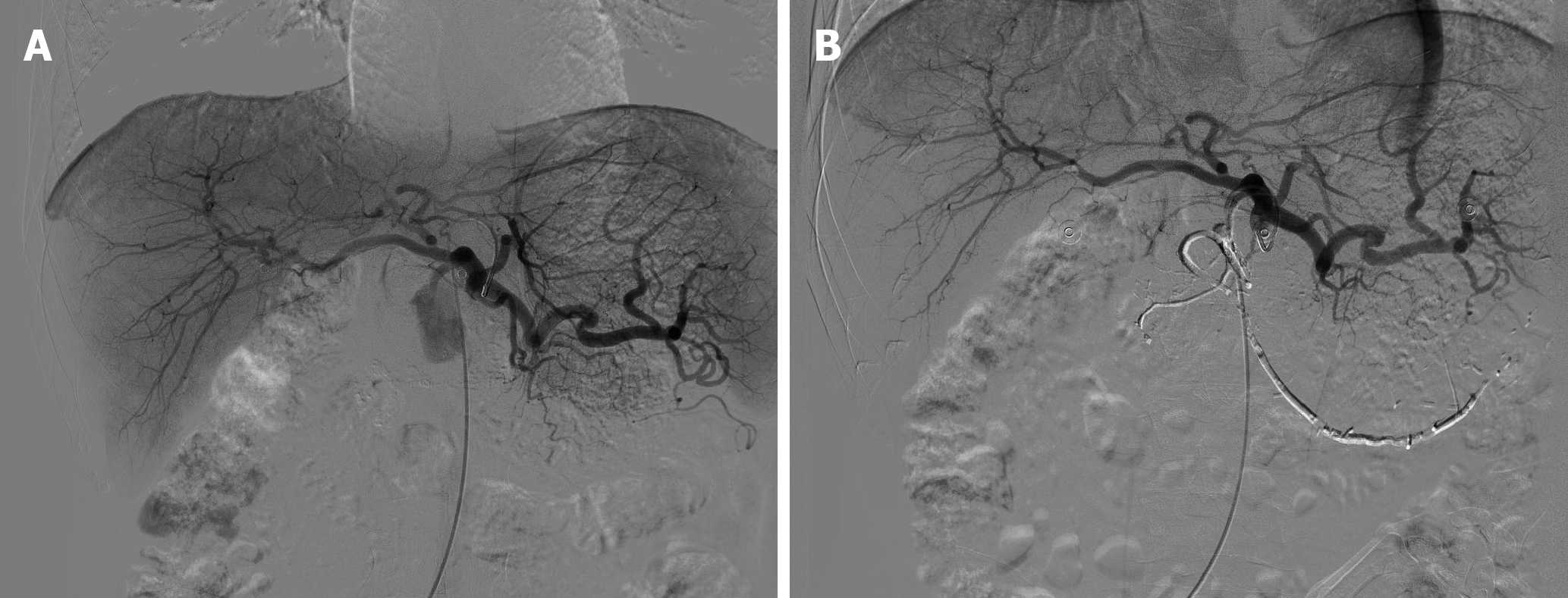
Figure 2 Abdominal digital subtraction angiography.
A: Gastroduodenal artery pseudoaneurysm; B: Image after occlusion using a coil and cyanoacrylate embolization.
- Citation: Cui HY, Jiang CH, Dong J, Wen Y, Chen YW. Hemosuccus pancreaticus caused by gastroduodenal artery pseudoaneurysm associated with chronic pancreatitis: A case report and review of literature. World J Clin Cases 2021; 9(1): 236-244
- URL: https://www.wjgnet.com/2307-8960/full/v9/i1/236.htm
- DOI: https://dx.doi.org/10.12998/wjcc.v9.i1.236


