Copyright
©The Author(s) 2020.
World J Clin Cases. Jan 6, 2021; 9(1): 204-210
Published online Jan 6, 2021. doi: 10.12998/wjcc.v9.i1.204
Published online Jan 6, 2021. doi: 10.12998/wjcc.v9.i1.204
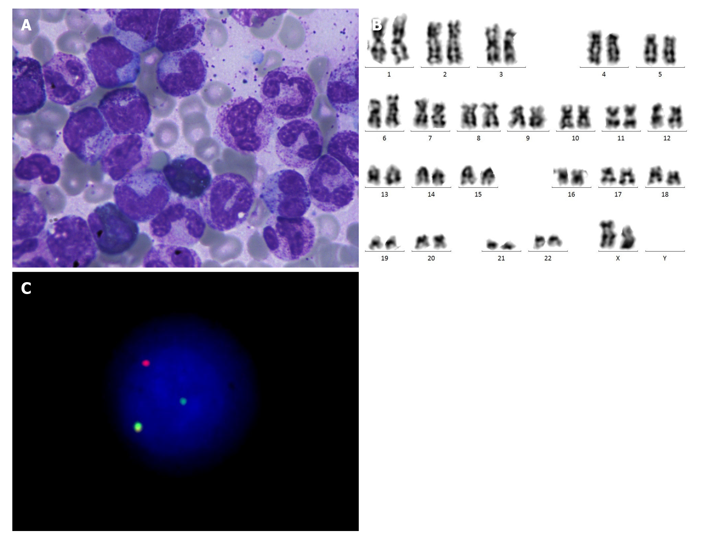
Figure 1 Myeloid neoplasm with eosinophilia and platelet-derived growth factor receptor beta rearrangement.
A: Myelomonocyte hyperplasia and eosinophilia (aspirate smears, Wright-Giemsa stain, 1000 ×); B: Normal karyotype with 46, XX; C: Fluorescence in situ hybridization revealed platelet-derived growth factor receptor beta rearrangement.
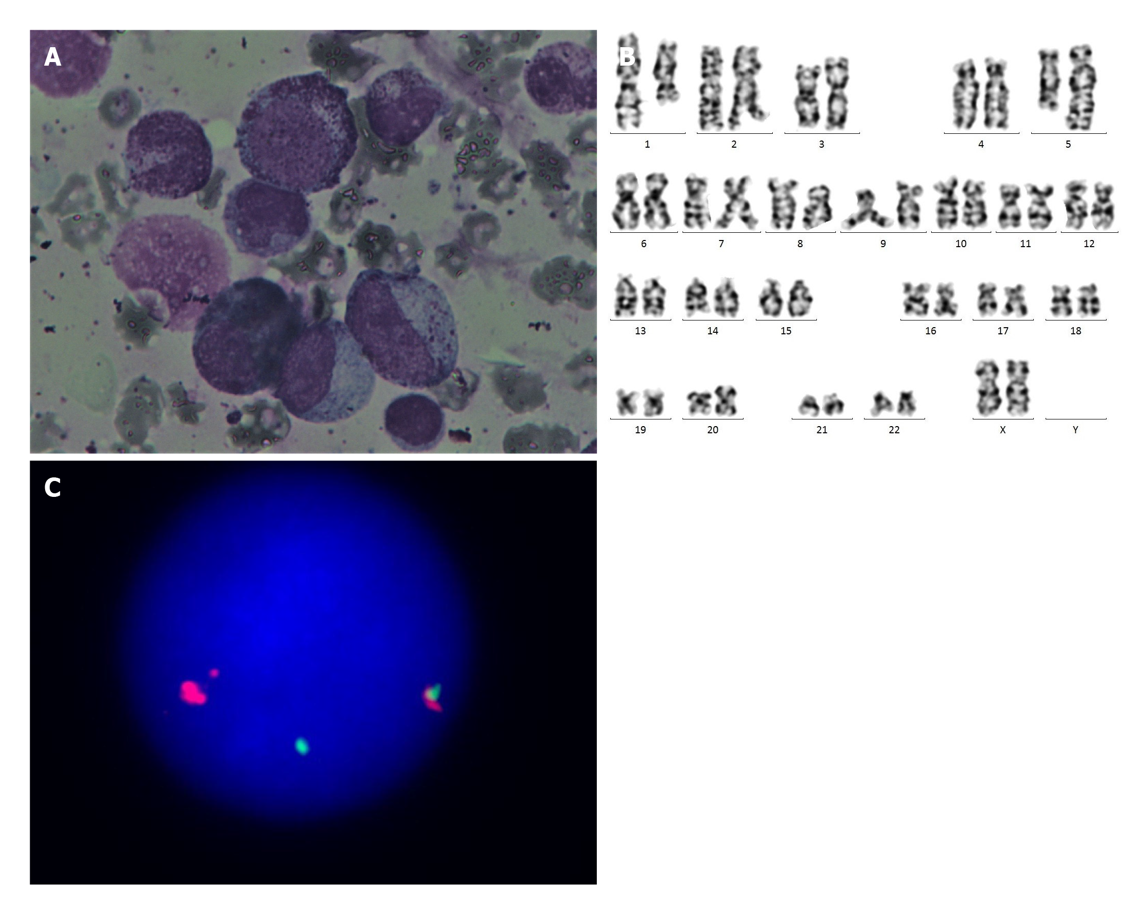
Figure 2 Myeloid neoplasm with eosinophilia and platelet-derived growth factor receptor beta rearrangement.
A: Myelomonocyte hyperplasia (aspirate smears, Wright-Giemsa stain, 1000 ×); B: Karyotype with 46, XX, t(1;5)(q21;q33); C: Fluorescence in situ hybridization revealed platelet-derived growth factor receptor beta rearrangement.
- Citation: Wang SC, Yang WY. Myeloid neoplasm with eosinophilia and rearrangement of platelet-derived growth factor receptor beta gene in children: Two case reports. World J Clin Cases 2021; 9(1): 204-210
- URL: https://www.wjgnet.com/2307-8960/full/v9/i1/204.htm
- DOI: https://dx.doi.org/10.12998/wjcc.v9.i1.204


