Copyright
©The Author(s) 2021.
World J Clin Cases. Jan 6, 2021; 9(1): 190-196
Published online Jan 6, 2021. doi: 10.12998/wjcc.v9.i1.190
Published online Jan 6, 2021. doi: 10.12998/wjcc.v9.i1.190
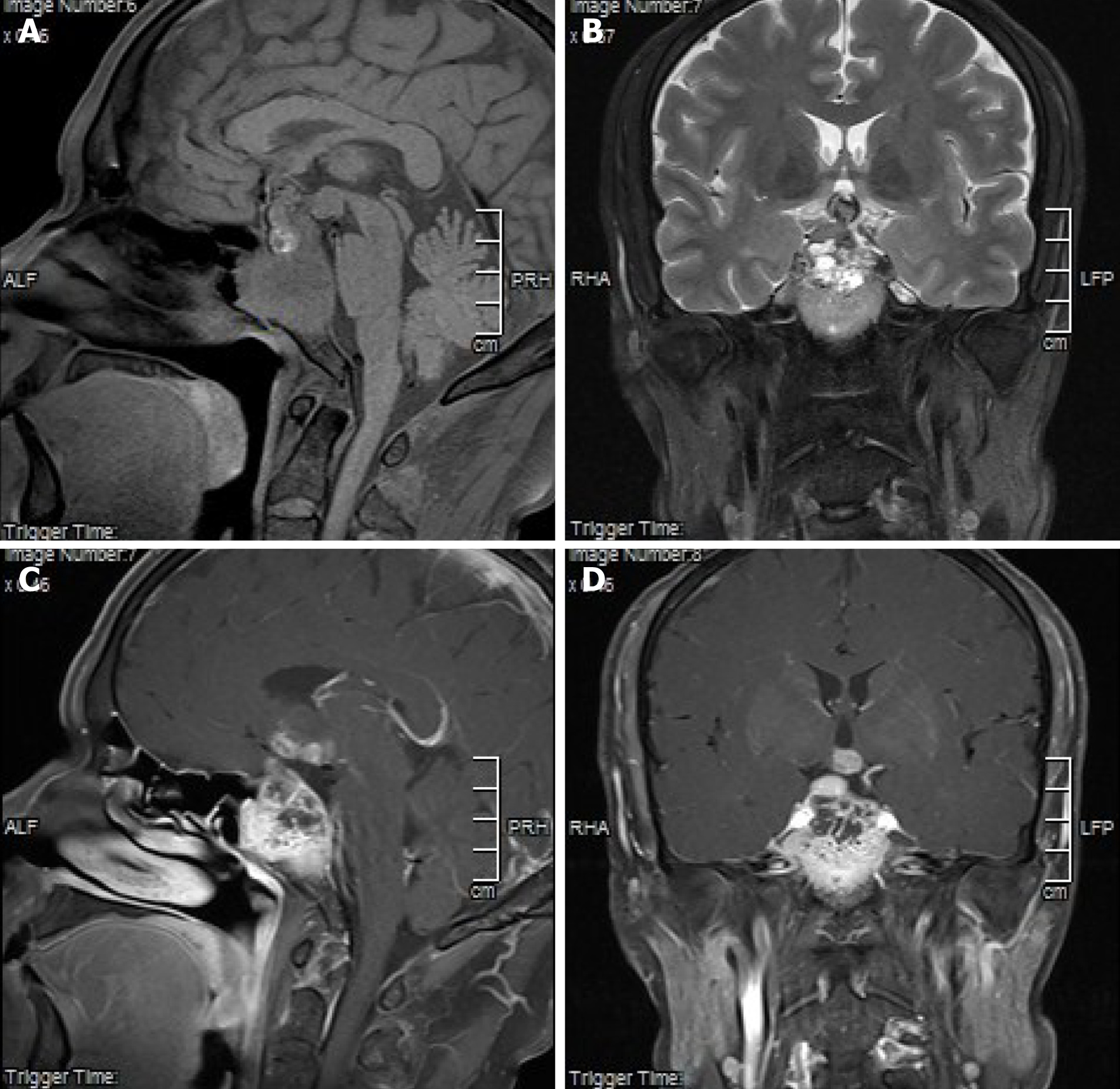
Figure 1 Imaging examination results.
A: T1-weighted images showed iso-and spot-like high- and low-intensity signals; B: T2-weighted images, which showed slightly high-, high-, and low-intensity signals; liquid-liquid plane; C and D: Inhomogeneous enhancement was seen in enhanced scan. Pituitary and pituitary stalk were not clearly visible. Optic nerve and papillary body were surrounded, protruding to the third ventricle, invading bilateral cavernous sinus and internal carotid artery, and only a small part of slope was shown.
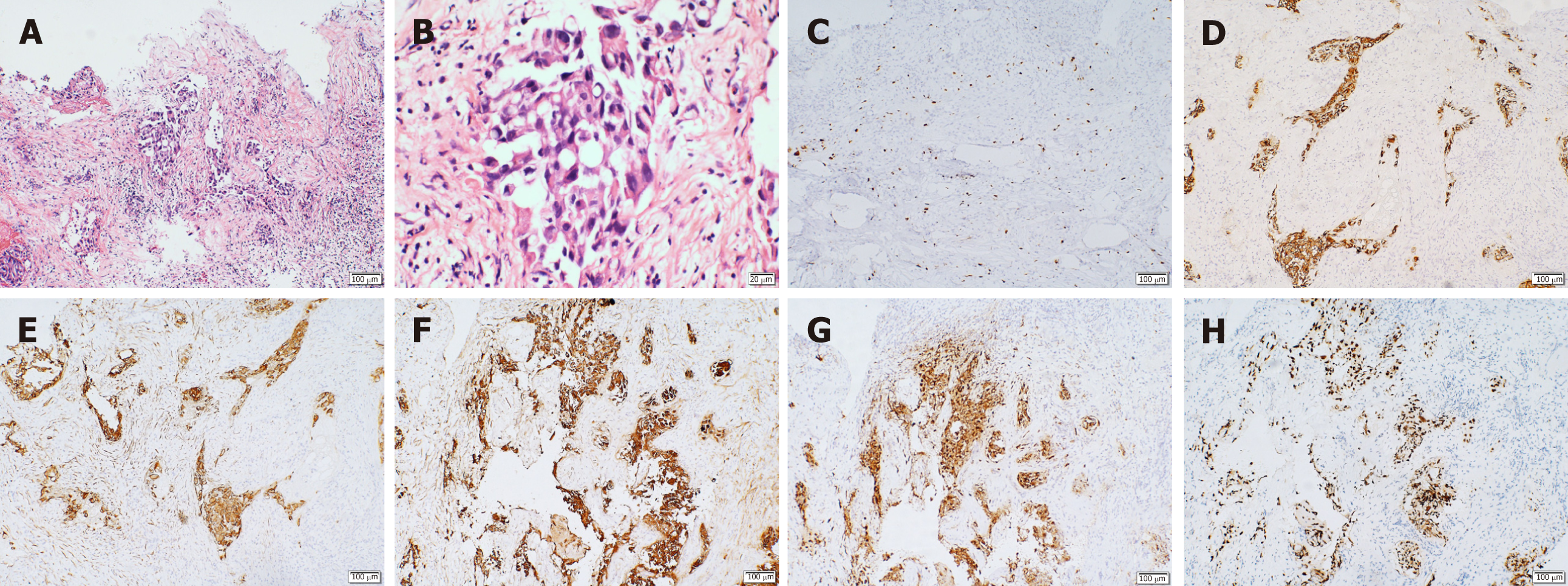
Figure 2 Immunohistochemistry results.
A: Increased nucleus and mitosis (hematoxylin and eosin × 10); B: Low differentiation and increased mucus secretion (positive hematoxylin and eosin × 40); C: Ki-67, suggestive of active cell proliferation and low degree of differentiation; D: Cytokeratin (CK)-positive, suggestive of epithelial cell differentiation, and thus, supporting the diagnosis of lung cancer metastasis; E: CK7 (+), supporting the diagnosis of lung cancer; F: CK-pan (+), suggesting that the tissue originated from epithelial cells; G: napsin A (+), suggestive of primary lung adenocarcinoma; H: thyroid transcription factor 1 (+), suggestive of non-small cell lung cancer.
- Citation: Liu CY, Wang YB, Zhu HQ, You JL, Liu Z, Zhang XF. Hyperprolactinemia due to pituitary metastasis: A case report. World J Clin Cases 2021; 9(1): 190-196
- URL: https://www.wjgnet.com/2307-8960/full/v9/i1/190.htm
- DOI: https://dx.doi.org/10.12998/wjcc.v9.i1.190


