Copyright
©The Author(s) 2021.
World J Clin Cases. Jan 6, 2021; 9(1): 183-189
Published online Jan 6, 2021. doi: 10.12998/wjcc.v9.i1.183
Published online Jan 6, 2021. doi: 10.12998/wjcc.v9.i1.183
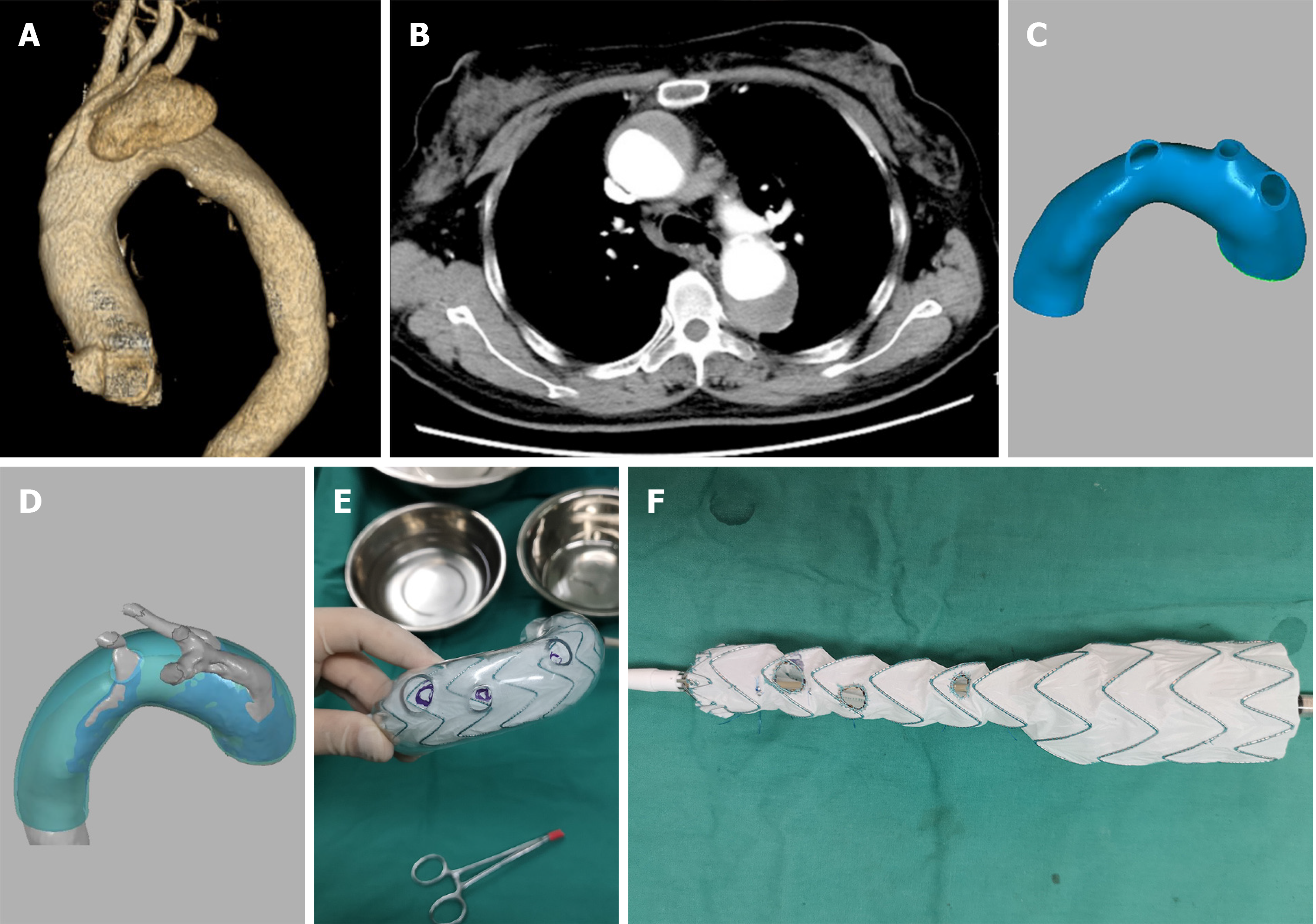
Figure 1 Details of treatment.
A and B: Preoperative computed tomography reconstruction and cross-sectional image; C and D: Design of the three-dimensional-printed model; E and F: Determination of the fenestrations and reduction in the diameter of the stent.
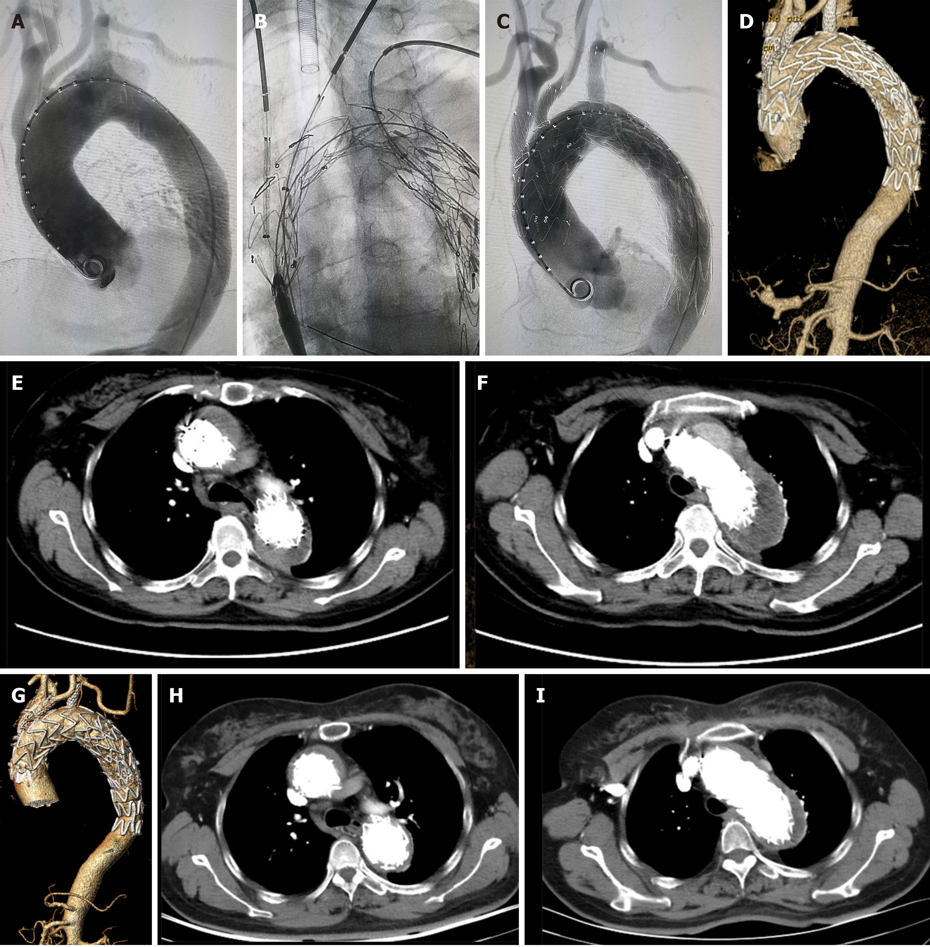
Figure 2 Postoperative follow-up.
A: Preoperative angiography; B: Three branched stents entering the aortic stent; C: Postoperative angiography of the aortic arch; D-F: Computed tomography reconstruction and cross-sectional image at 3 mo after surgery; G and I: Computed tomography reconstruction and cross-sectional image at 1 year after surgery.
- Citation: Zhang M, Tong YH, Liu C, Li XQ, Liu CJ, Liu Z. Treatment of Stanford type A aortic dissection with triple pre-fenestration, reduced diameter, and three-dimensional-printing techniques: A case report. World J Clin Cases 2021; 9(1): 183-189
- URL: https://www.wjgnet.com/2307-8960/full/v9/i1/183.htm
- DOI: https://dx.doi.org/10.12998/wjcc.v9.i1.183


