Copyright
©The Author(s) 2021.
World J Clin Cases. Jan 6, 2021; 9(1): 175-182
Published online Jan 6, 2021. doi: 10.12998/wjcc.v9.i1.175
Published online Jan 6, 2021. doi: 10.12998/wjcc.v9.i1.175
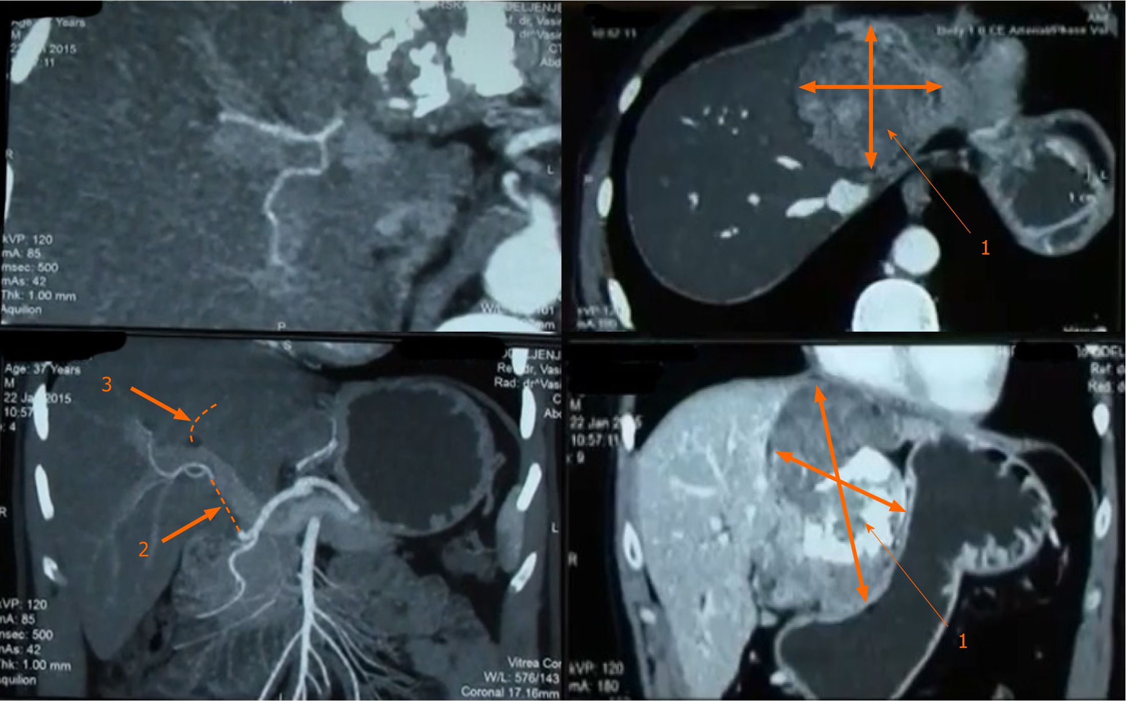
Figure 1 Multi-detector computed tomography of the abdomen.
1: Tumor; 2: Infiltration of proper hepatic artery from the origin of the gastro-duodenal aretry up to the right second branching 3-complete infiltration of the left portal vein.
Figure 2 Histopathology.
A: Mix of heavily collagenizedhypocellular zones -giant rosettes and cell-rich part of tumor [hematoxylin eosin staining (HE), 40 ×]; B: Short fascicular and characteristic whorling growth patterns are often seen. There are arcades of curvilinear blood vessels accompanied by perivascular hyaline degeneration (HE, 100 ×).
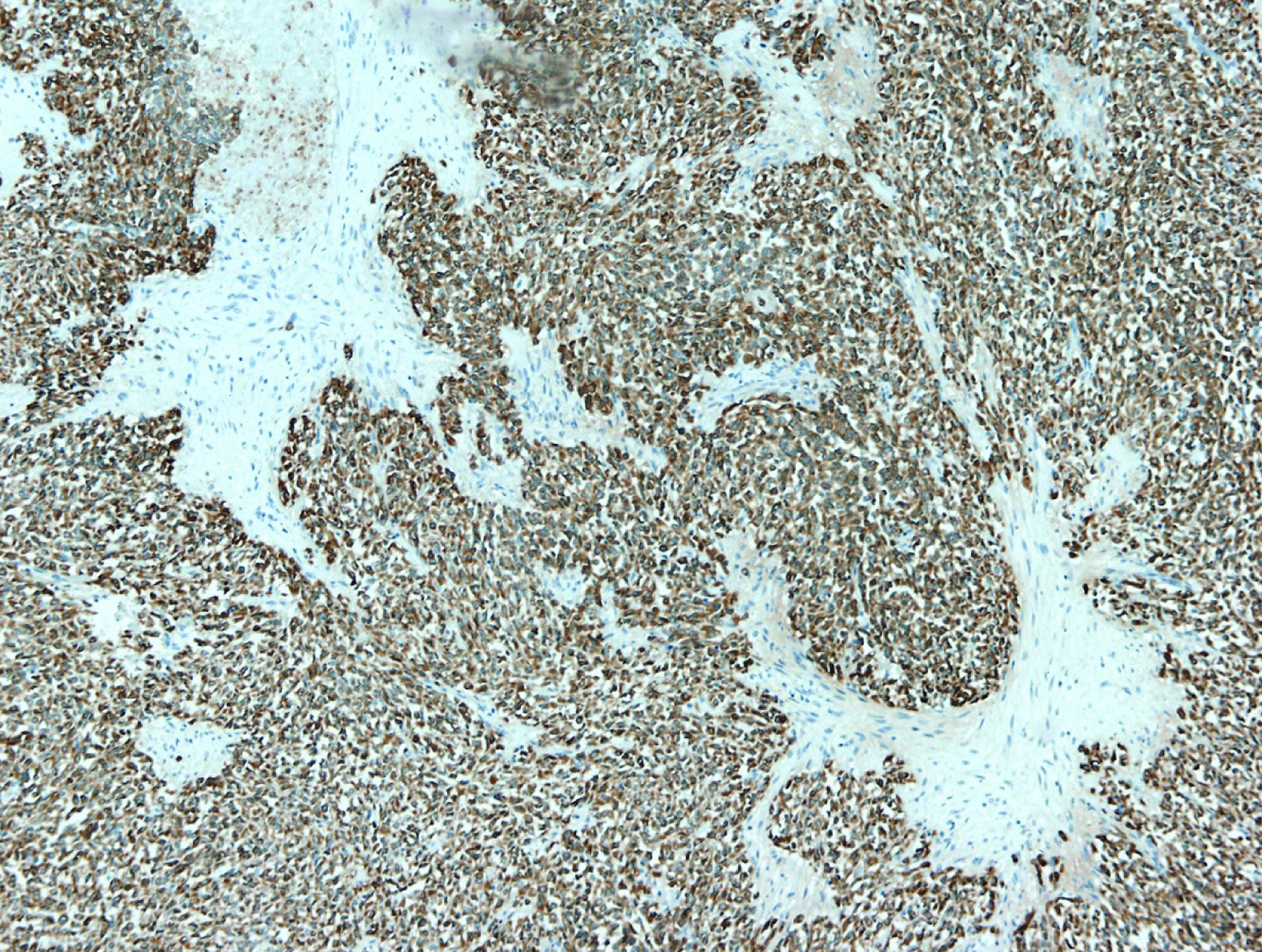
Figure 3 Immunohistochemistry.
Tumor cells were diffusely and strongly positive for vimentin and MUC4, CD99 and epithelial membrane antigen were diffuse and slight expressed, and cytokeratin, smooth muscle actin, S-100 protein and neuron specific enolase were negative.
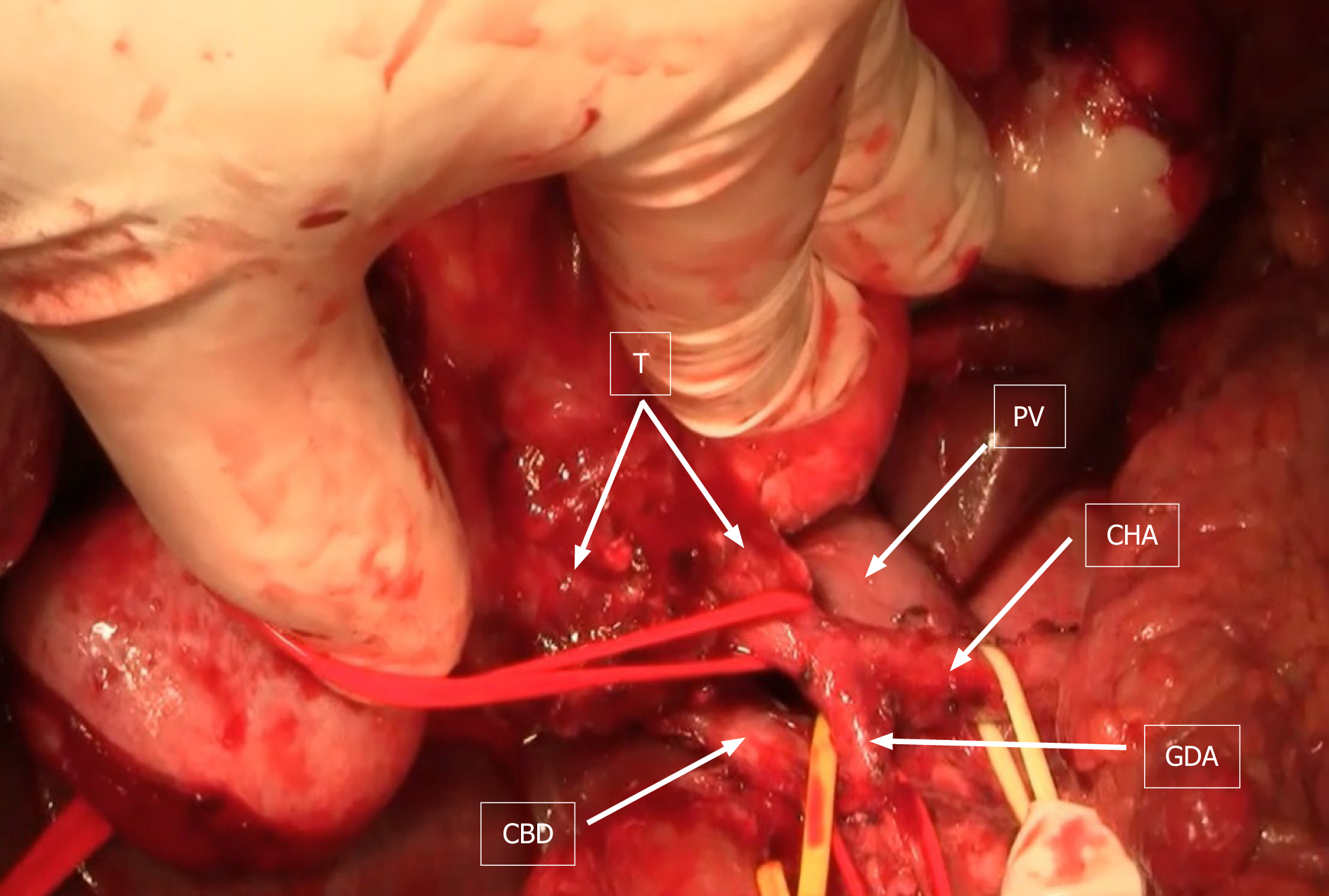
Figure 4 Intraoperative finding.
T: Tumor; PV: Portal vein; CHA: Common hepatic artery; GDA: Gastroduodenal artery; CBD: Common bile duct.
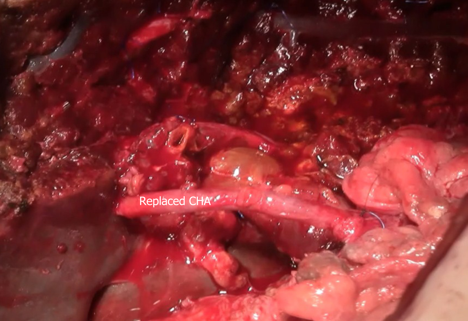
Figure 5 Reconstruction of the common hepatic artery with saphenous vein graft.
CHA: Common hepatic artery.
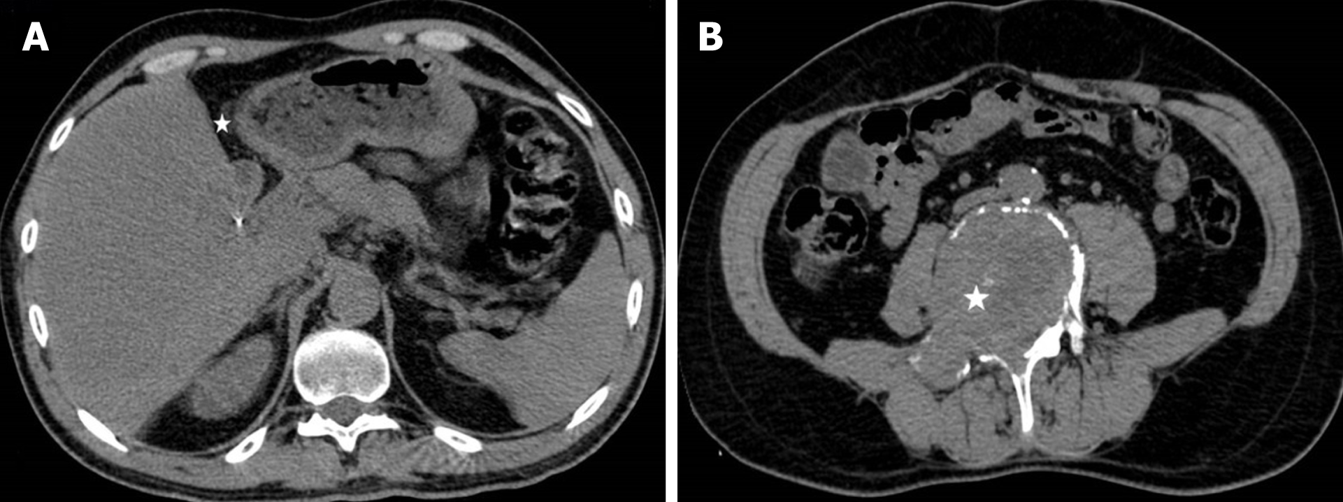
Figure 6 Abdominal and pelvic computed tomography after left hepatectomy-axial images.
A: There is no tumor reccurence on surgical margin (white star) and no focal lesions in right liver lobe; B: An ill-defined lytic lesion (white star) of the L5 vertebral body is seen without periosteal reaction, representing solitary osseous metastasis of liver sarcoma.
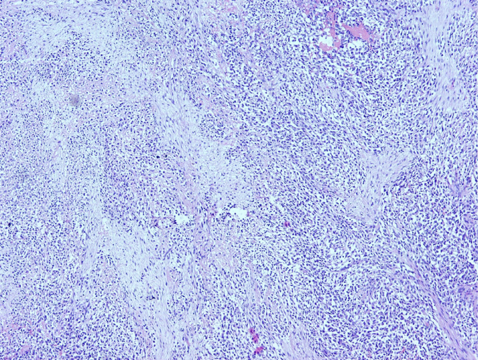
Figure 7 Histopathology.
Prominent vascularity in myxoid areas and perivascular hypercellularity seen in metastatic tumor corelate with changes in primary tumor.
- Citation: Dugalic V, Ignjatovic II, Kovac JD, Ilic N, Sopta J, Ostojic SR, Vasin D, Bogdanovic MD, Dumic I, Milovanovic T. Low-grade fibromyxoid sarcoma of the liver: A case report . World J Clin Cases 2021; 9(1): 175-182
- URL: https://www.wjgnet.com/2307-8960/full/v9/i1/175.htm
- DOI: https://dx.doi.org/10.12998/wjcc.v9.i1.175


