Copyright
©The Author(s) 2020.
World J Clin Cases. May 6, 2020; 8(9): 1752-1755
Published online May 6, 2020. doi: 10.12998/wjcc.v8.i9.1752
Published online May 6, 2020. doi: 10.12998/wjcc.v8.i9.1752
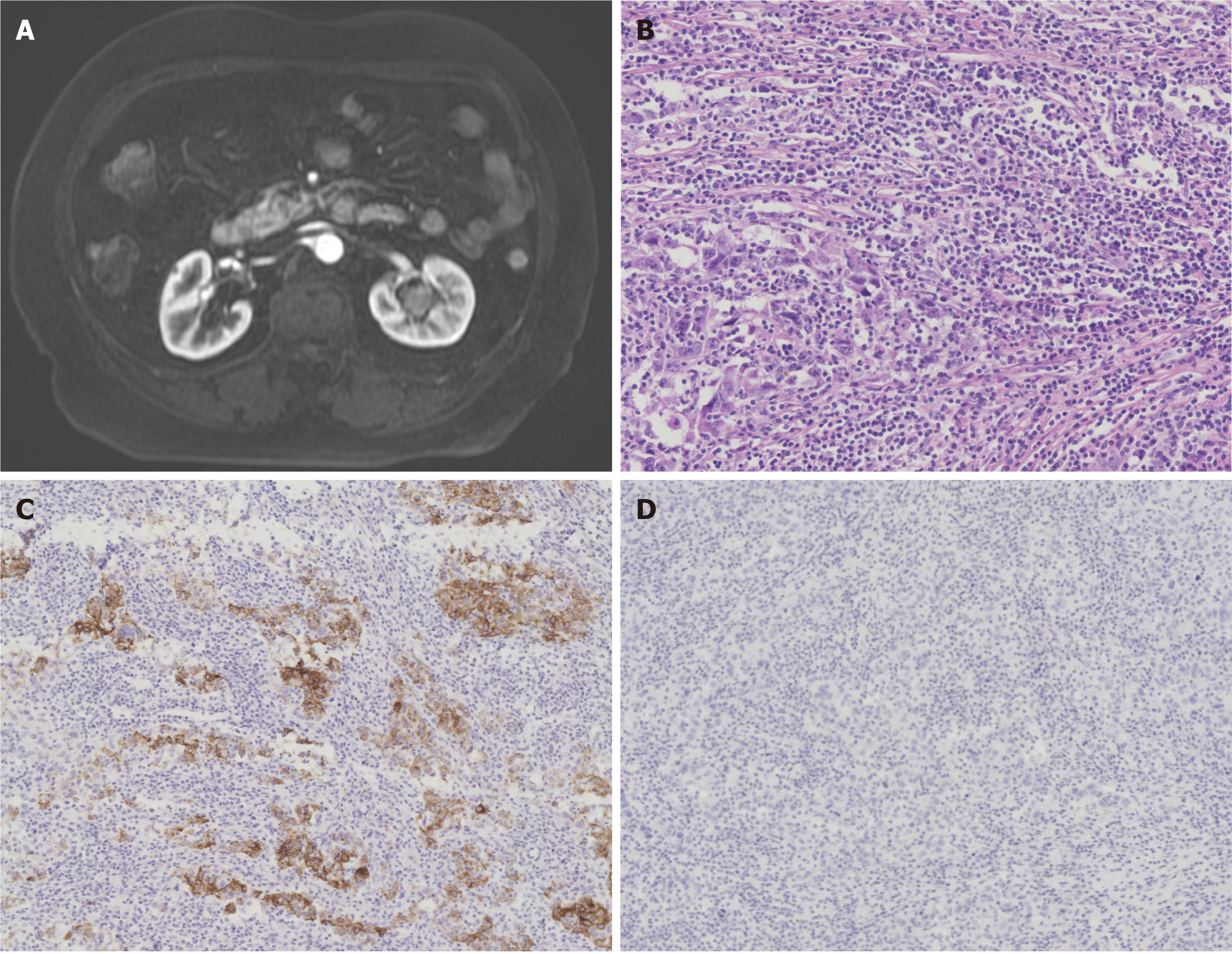
Figure 1 Clinical imaging and pathological features.
A: Magnetic resonance imaging showing a slightly enhancing lesion within the left renal pelvis; B: Haematoxylin and eosin staining showing abundant lymphoid stroma surrounding the large polygonal tumour cells; C: Diffuse cytokeratin 7 immunoreactivity highlighting the epithelial component of the tumour; D: Immunohistochemical staining showing tumour cell without Epstein-Barr virus present.
- Citation: Lai SC, Seery S, Diao TX, Wang JY, Liu M. Rare primary lymphoepithelioma-like carcinoma of the renal pelvis. World J Clin Cases 2020; 8(9): 1752-1755
- URL: https://www.wjgnet.com/2307-8960/full/v8/i9/1752.htm
- DOI: https://dx.doi.org/10.12998/wjcc.v8.i9.1752


