Copyright
©The Author(s) 2020.
World J Clin Cases. May 6, 2020; 8(9): 1721-1728
Published online May 6, 2020. doi: 10.12998/wjcc.v8.i9.1721
Published online May 6, 2020. doi: 10.12998/wjcc.v8.i9.1721
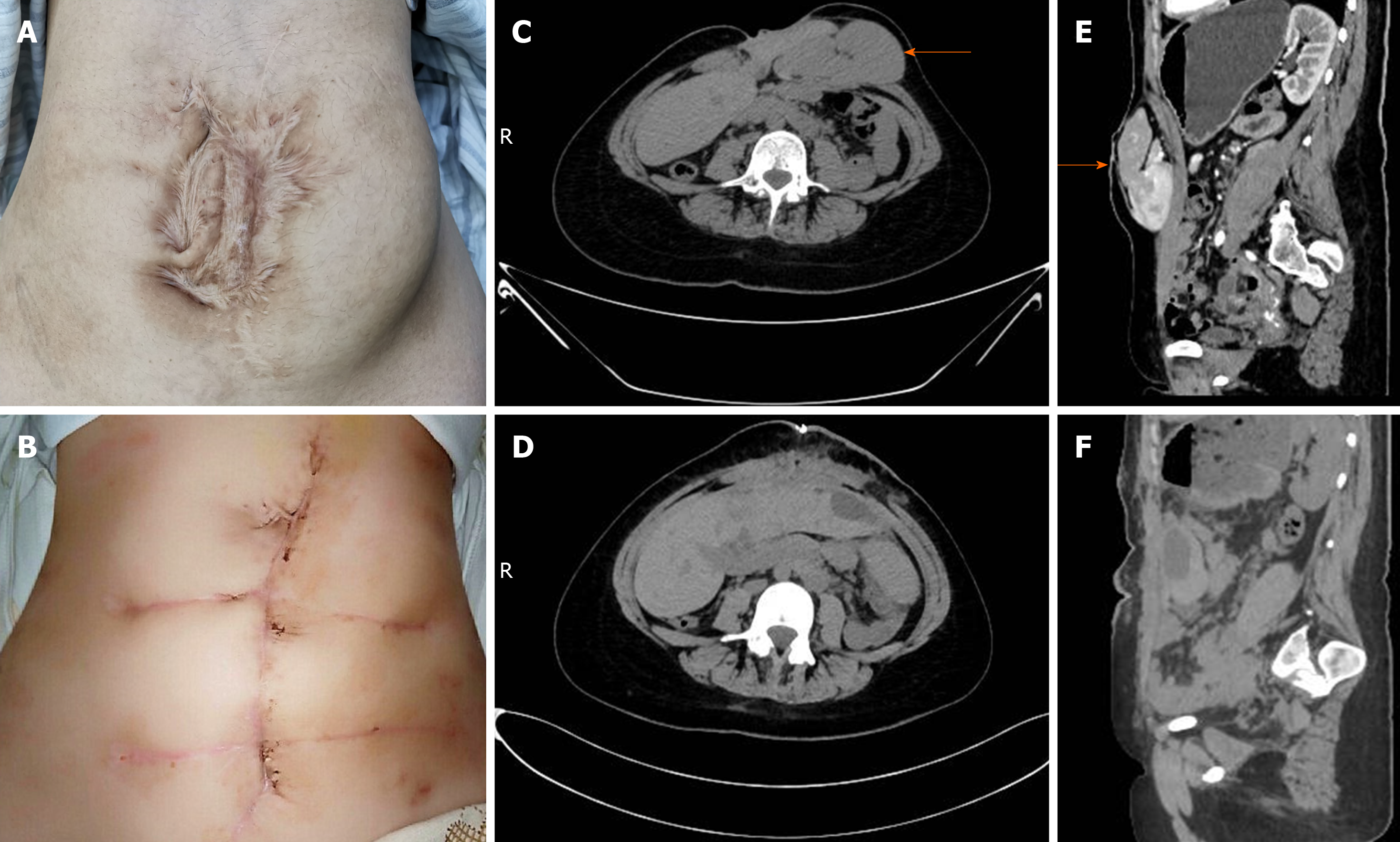
Figure 1 Patient-related image.
A: Many irregular and obsolete surgical scars on the abdominal wall; B: The appearance of the surgical incision improved greatly after operation; C and E: Computed tomography demonstrated a giant ventral hernia containing liver, pancreas, spleen and blood vessels before operation (arrow); D and F: Liver, pancreas, spleen and blood vessels returned to the abdominal cavity after operation.
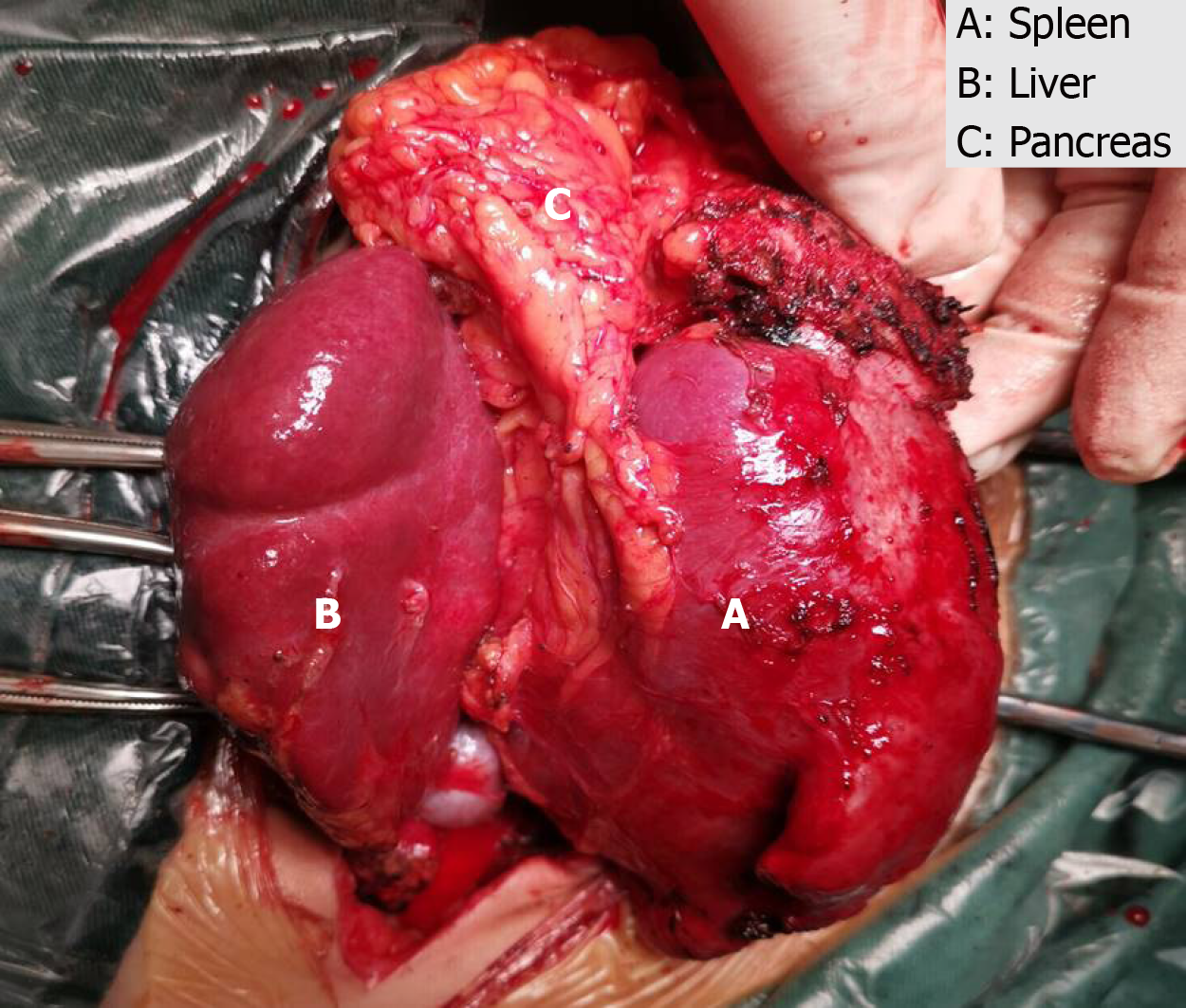
Figure 2 The spleen, part of the left lobe of the liver and the pancreas were observed between the adipose layer of the abdominal wall and the anterior sheath of the rectus abdominis.
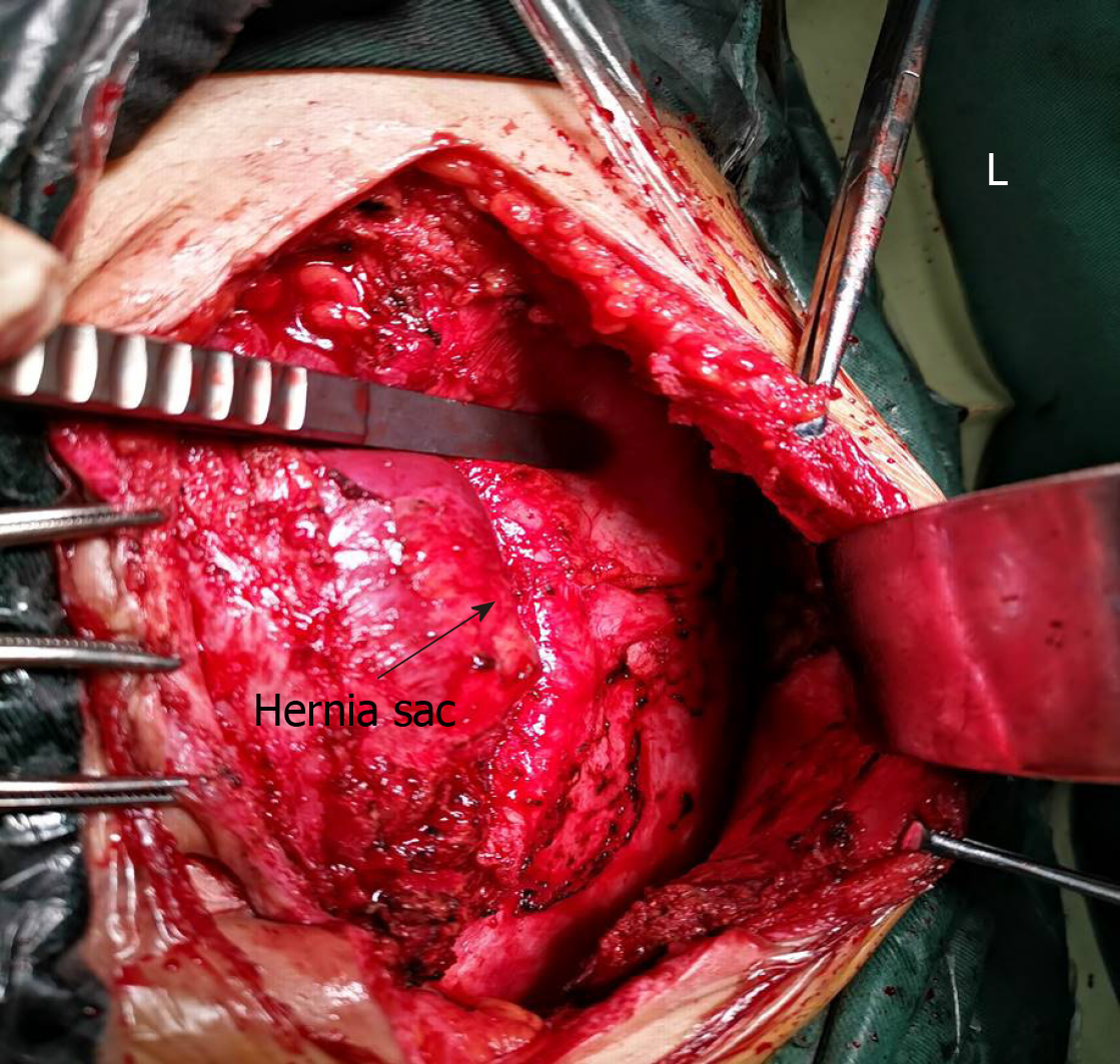
Figure 3 Approximately 3/4 of the hernia sac was located under the left abdominal wall (arrow) and 1/4 under the right abdominal wall.
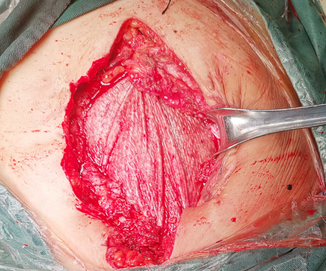
Figure 4 A COOK 15 cm × 13 cm biological Mesh was fixed to the retroperitoneum.
- Citation: Luo XG, Lu C, Wang WL, Zhou F, Yu CZ. Giant ventral hernia simultaneously containing the spleen, a portion of the pancreas and the left hepatic lobe: A case report. World J Clin Cases 2020; 8(9): 1721-1728
- URL: https://www.wjgnet.com/2307-8960/full/v8/i9/1721.htm
- DOI: https://dx.doi.org/10.12998/wjcc.v8.i9.1721


