Copyright
©The Author(s) 2020.
World J Clin Cases. May 6, 2020; 8(9): 1698-1704
Published online May 6, 2020. doi: 10.12998/wjcc.v8.i9.1698
Published online May 6, 2020. doi: 10.12998/wjcc.v8.i9.1698
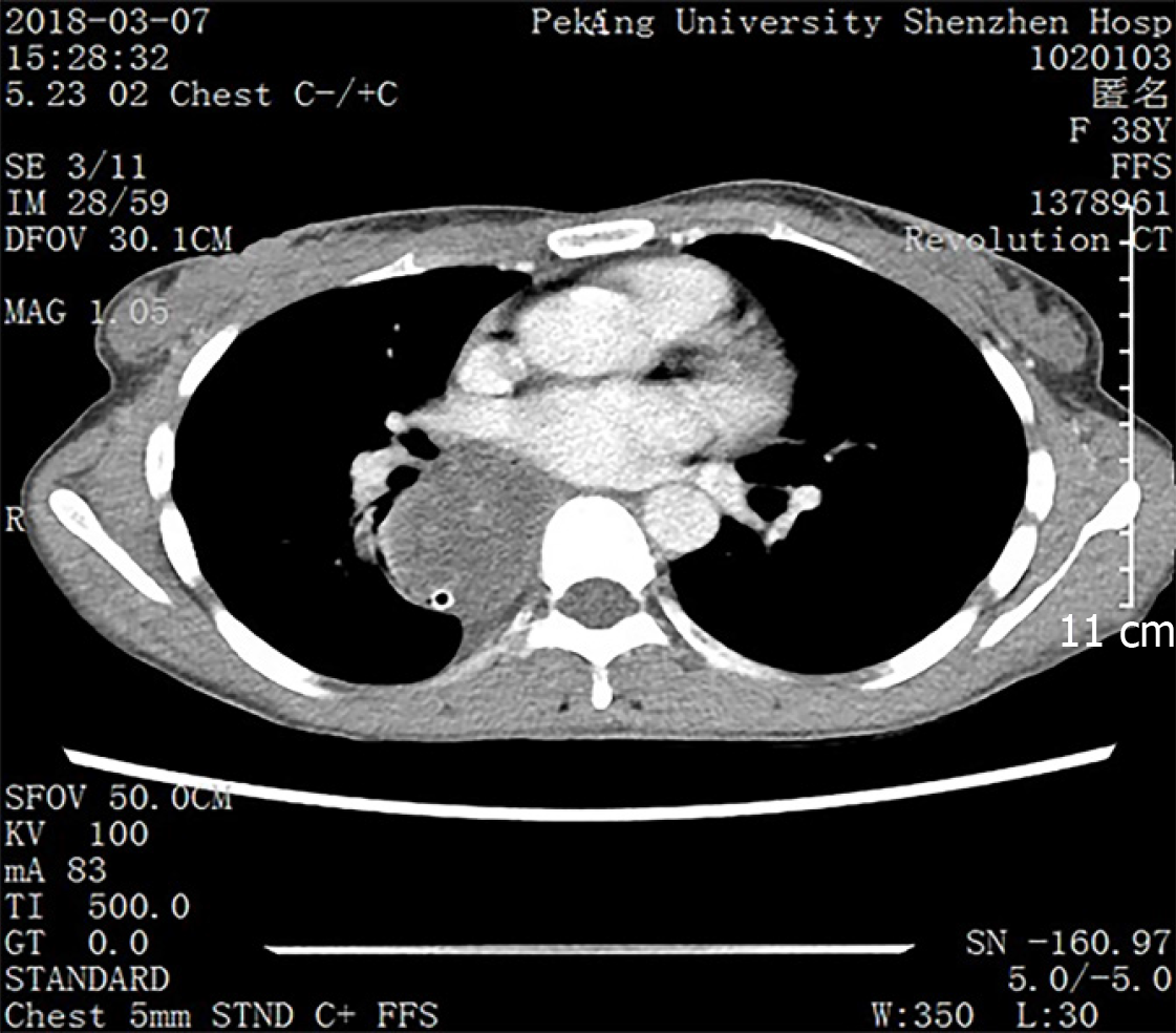
Figure 1 Computed tomography showed an expanded esophagus and low density shadow.
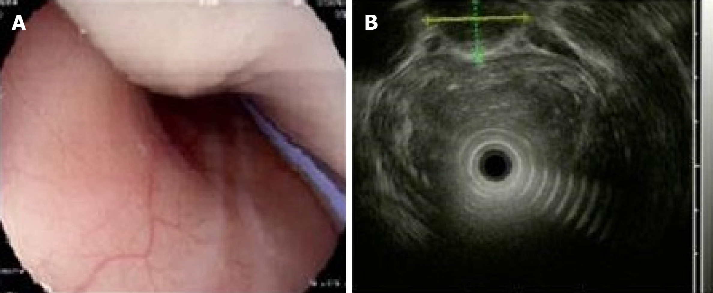
Figure 2 Esophagogastric ultrasonography showed a homogeneous, low echogenic mass outside the esophagus.
A: Esophagogastroscopy showed a large mass pushing against the esophagus; B: Esophagogastric ultrasonography showed a large, homogeneous, low echogenic mass outside the esophagus, 20–40 cm below the incisor, with a cross-sectional area of 21.5 mm × 15.9 mm.
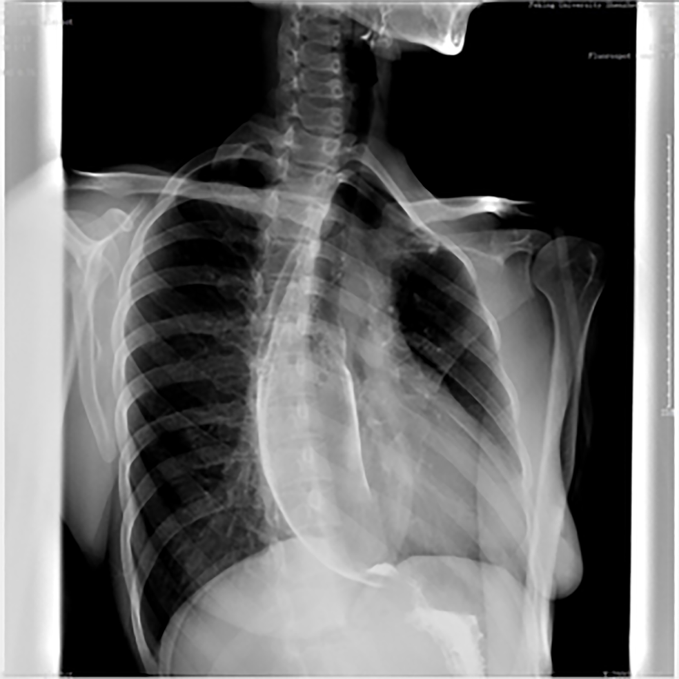
Figure 3 Upper gastrointestinal angiography showed external pressure stenosis of the esophagus.
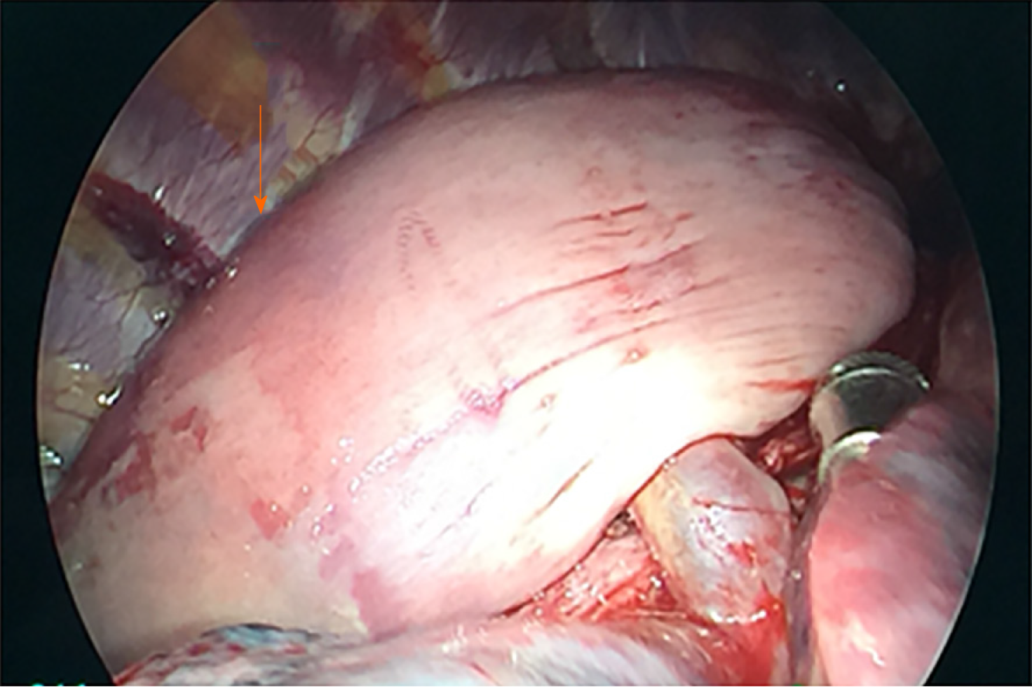
Figure 4 Intraoperative photograph showing a large, smooth-surface mass (orange arrow) in the posterior mediastinum and the esophageal muscle layer was intact.
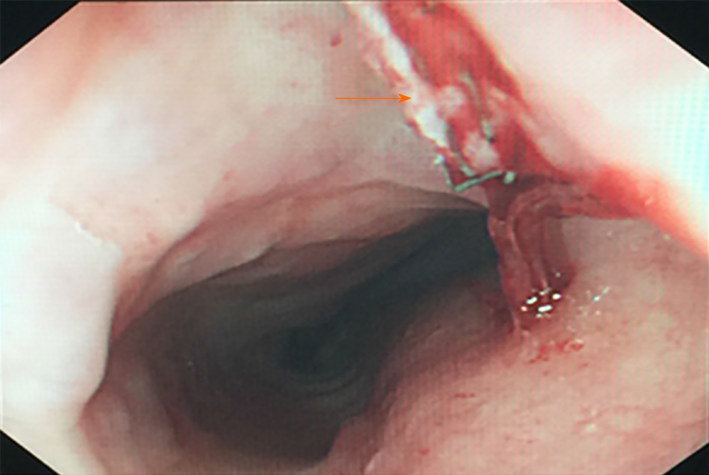
Figure 5 Little residual mass was seen by gastroscopy.
The direction of the orange arrow is the incisal margin.
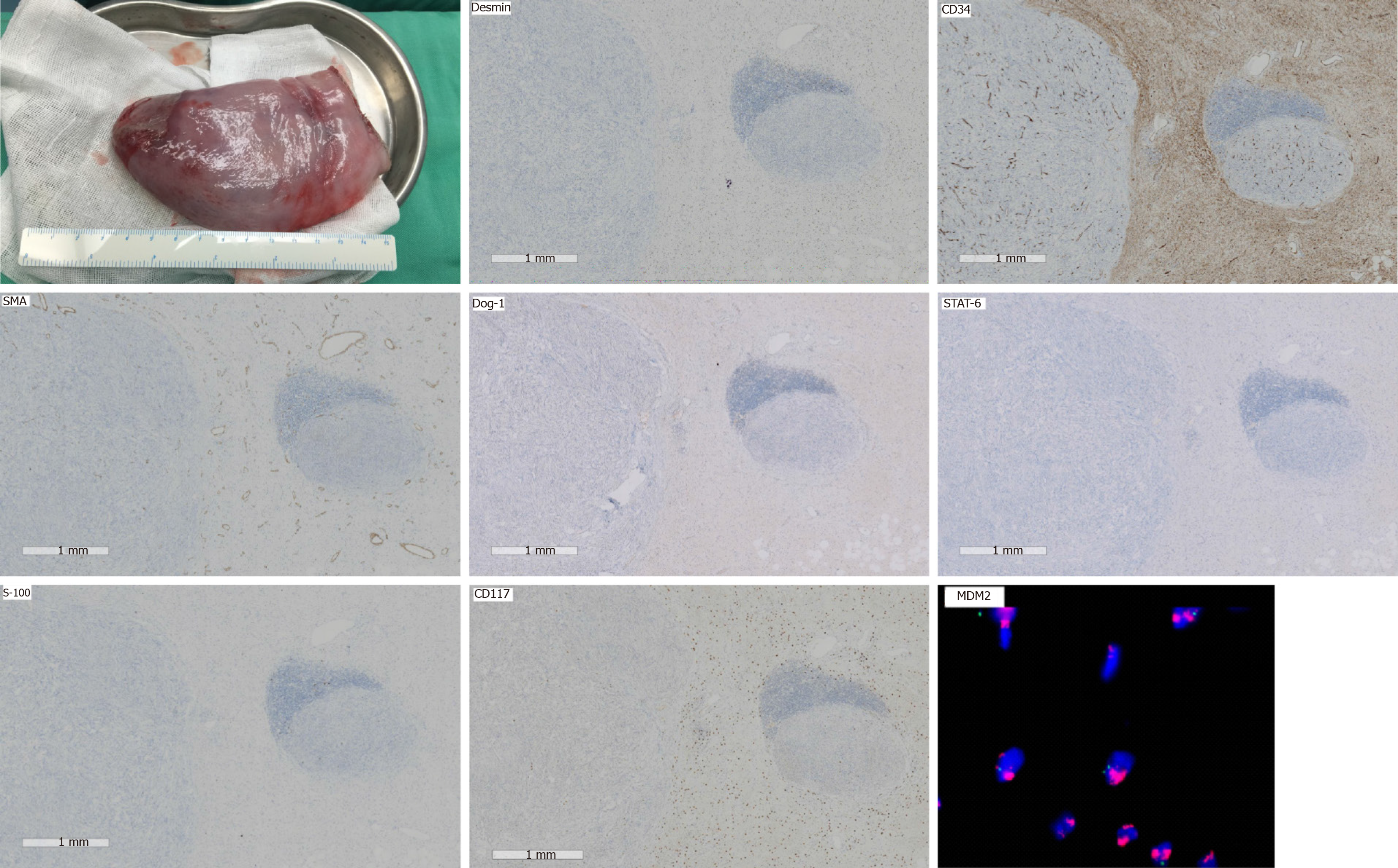
Figure 6 Immunohistochemical analysis revealed some discrete spindle cells which were positive for Desmin and CD34, and negative for S-100, SMA, DOG-1 and STAT-6.
Fatty tissue was positive for S-100 protein. CD117 highlighted the intermixed mast cells and plasma cells. MDM2 gene amplification was confirmed by fluorescence in situ hybridization analysis, and each one is marked in red in the upper right corner.
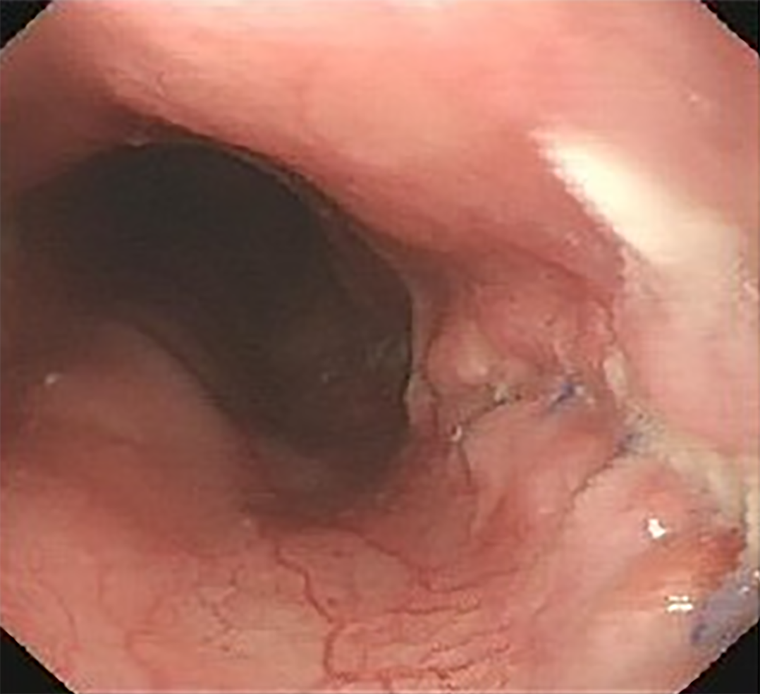
Figure 7 Esophagoscopy did not find any mass in the esophageal lumen 20 mo after surgery.
- Citation: Ye YW, Liao MY, Mou ZM, Shi XX, Xie YC. Thoracoscopic resection of a huge esophageal dedifferentiated liposarcoma: A case report. World J Clin Cases 2020; 8(9): 1698-1704
- URL: https://www.wjgnet.com/2307-8960/full/v8/i9/1698.htm
- DOI: https://dx.doi.org/10.12998/wjcc.v8.i9.1698


