Copyright
©The Author(s) 2020.
World J Clin Cases. May 6, 2020; 8(9): 1679-1684
Published online May 6, 2020. doi: 10.12998/wjcc.v8.i9.1679
Published online May 6, 2020. doi: 10.12998/wjcc.v8.i9.1679
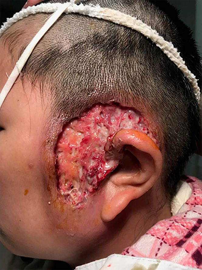
Figure 1 Physical examination of the left face.
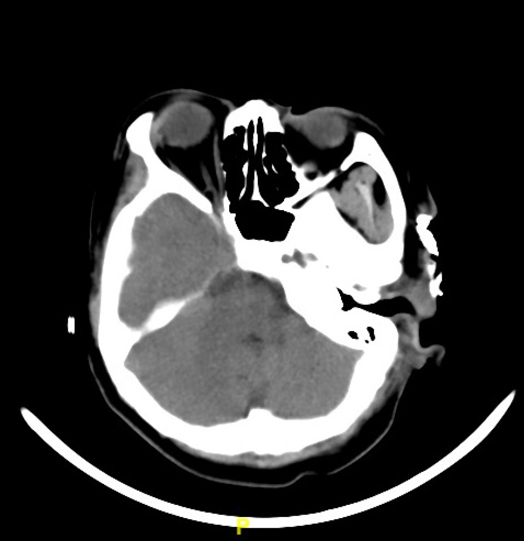
Figure 2 Axial CT scan.
Skin and subdermal soft-tissue defect without obvious erosion into the facial muscle, parotid gland and external auditory canal; high-density material lateral to zygomatic arch is iodoform gauze.
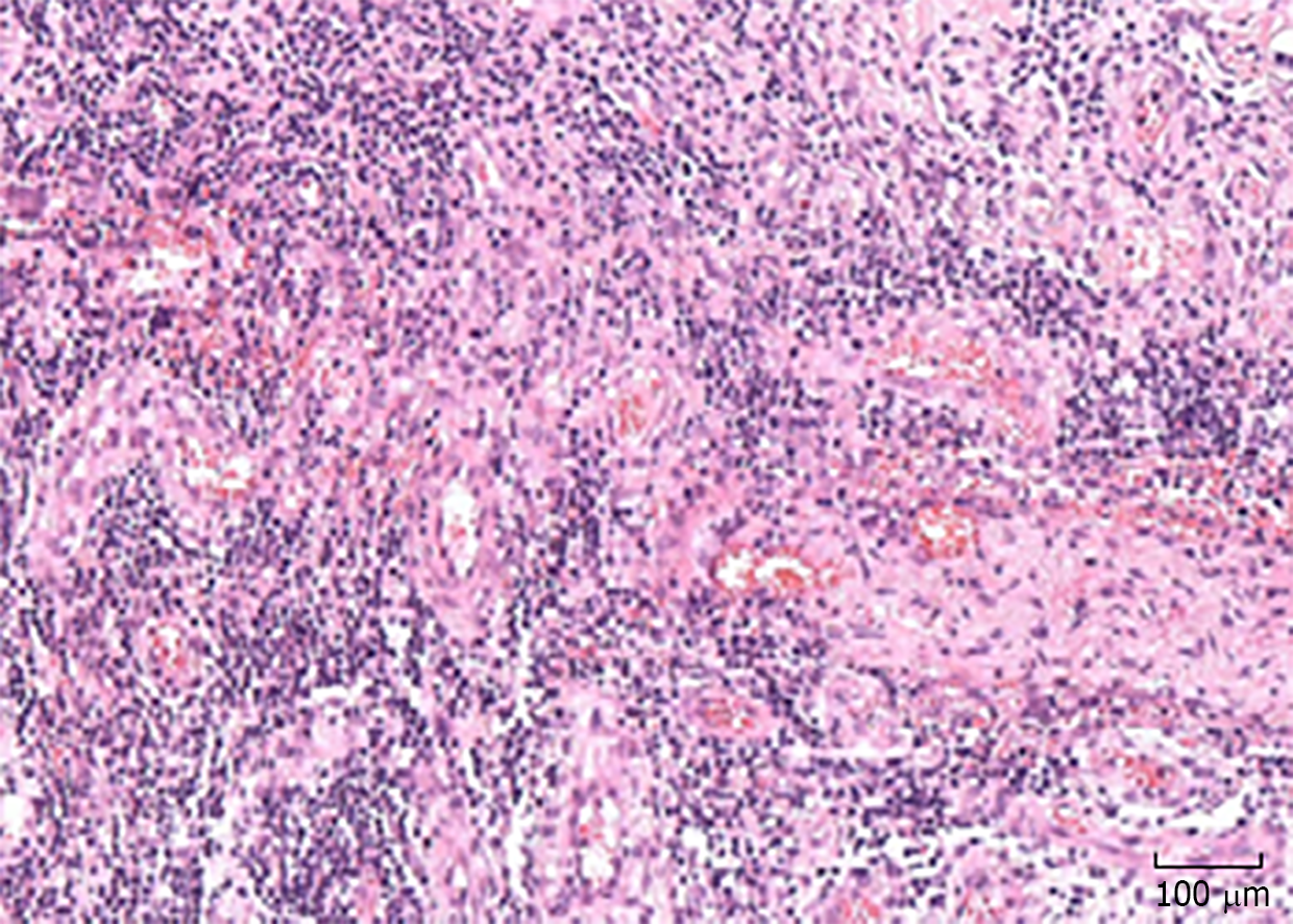
Figure 3 Biopsy specimen: widespread infiltration of inflammatory cells.
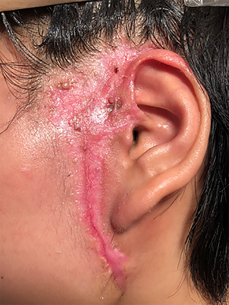
Figure 4 Appearance of auricular area after 6 mo.
The preauricular sinus is still visible.
- Citation: Zhao Y, Fang RY, Feng GD, Cui TT, Gao ZQ. Pyoderma gangrenosum confused with congenital preauricular fistula infection: A case report. World J Clin Cases 2020; 8(9): 1679-1684
- URL: https://www.wjgnet.com/2307-8960/full/v8/i9/1679.htm
- DOI: https://dx.doi.org/10.12998/wjcc.v8.i9.1679


