Copyright
©The Author(s) 2020.
World J Clin Cases. Apr 6, 2020; 8(7): 1306-1310
Published online Apr 6, 2020. doi: 10.12998/wjcc.v8.i7.1306
Published online Apr 6, 2020. doi: 10.12998/wjcc.v8.i7.1306
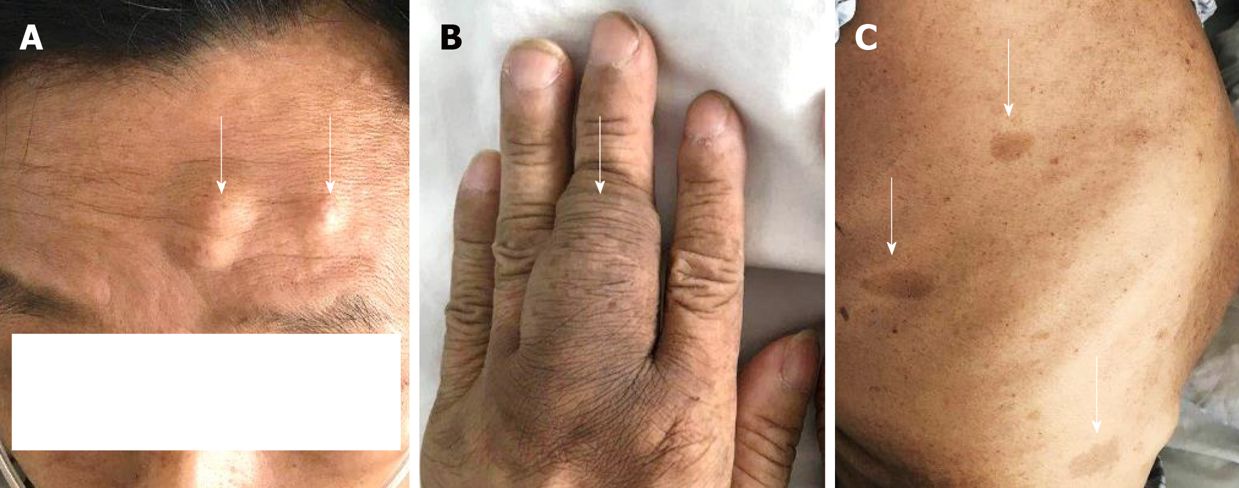
Figure 1 Multiple subcutaneous nodules throughout the body.
A: Two subcutaneous nodules in the forehead; B: Subcutaneous nodule on the middle phalanx of the left hand; C: Pigmentation on the right hypochondrium of the torso.
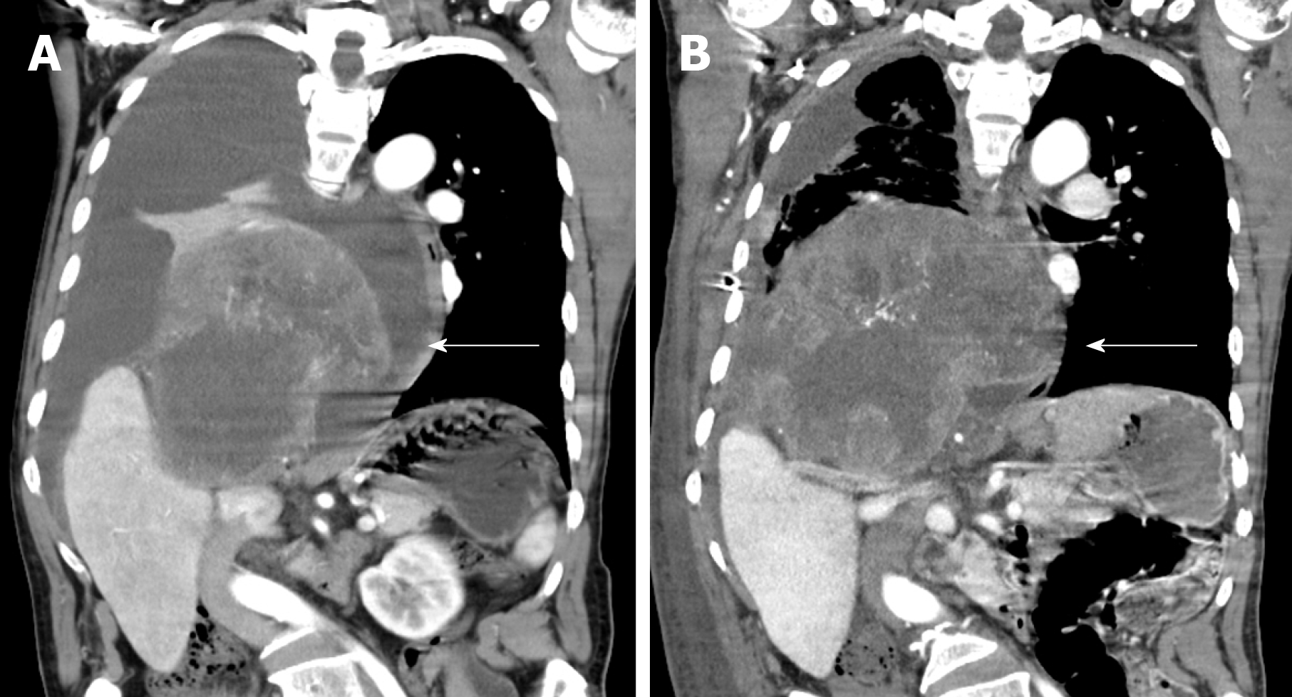
Figure 2 Changes in the patient’s tumor size from day 1 to day 24 after hospitalization.
A: A large mass in the right thoracic cavity, accompanied by extensive hydrothorax on the same side; B: Tumor growth in the right thoracic cavity was obvious, while the hydrothorax was significantly reduced after treatment.
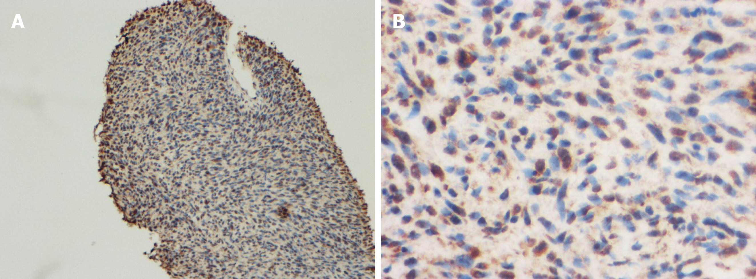
Figure 3 Microscopic findings show neurofibrosarcoma.
A: Abnormal hyperplasia of high chromatin cells (immunohistochemistry, × 100); B: Abnormally proliferating spindle cells (immunohistochemistry, × 400).
- Citation: Wang Y, Lu XF, Chen LL, Zhang YW, Zhang B. Multiple neurofibromas plus fibrosarcoma with familial NF1 pathogenicity: A case report. World J Clin Cases 2020; 8(7): 1306-1310
- URL: https://www.wjgnet.com/2307-8960/full/v8/i7/1306.htm
- DOI: https://dx.doi.org/10.12998/wjcc.v8.i7.1306


