Copyright
©The Author(s) 2020.
World J Clin Cases. Apr 6, 2020; 8(7): 1287-1294
Published online Apr 6, 2020. doi: 10.12998/wjcc.v8.i7.1287
Published online Apr 6, 2020. doi: 10.12998/wjcc.v8.i7.1287
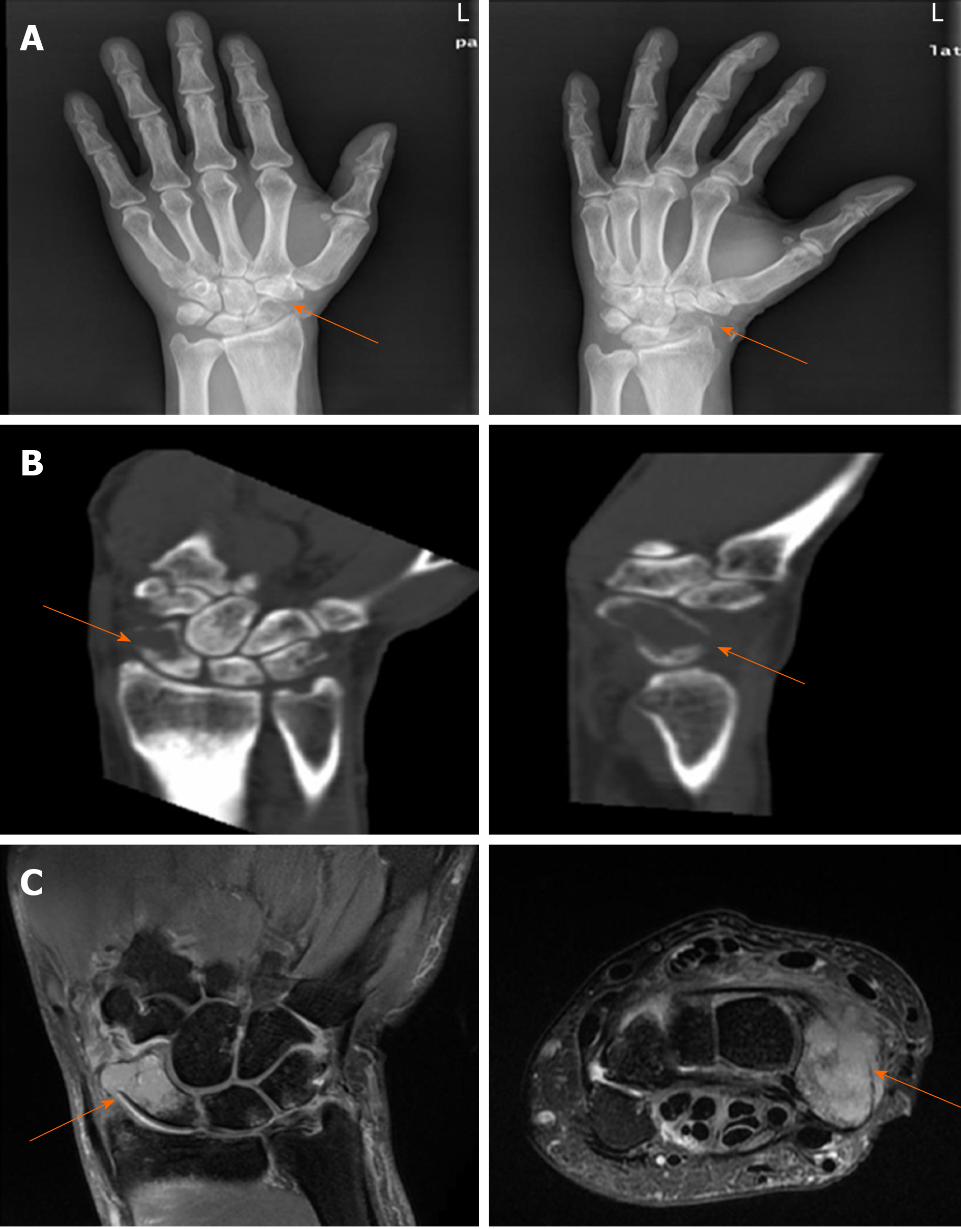
Figure 1 Imaging findings in the left hand.
A: Plain X-ray showed a decrease in scaphoid bone density accompanied by a fracture; B and C: Computed tomography and magnetic resonance imaging scans demonstrated scaphoid and triangular bone destruction and soft tissue swelling, indicating a pathological fracture.
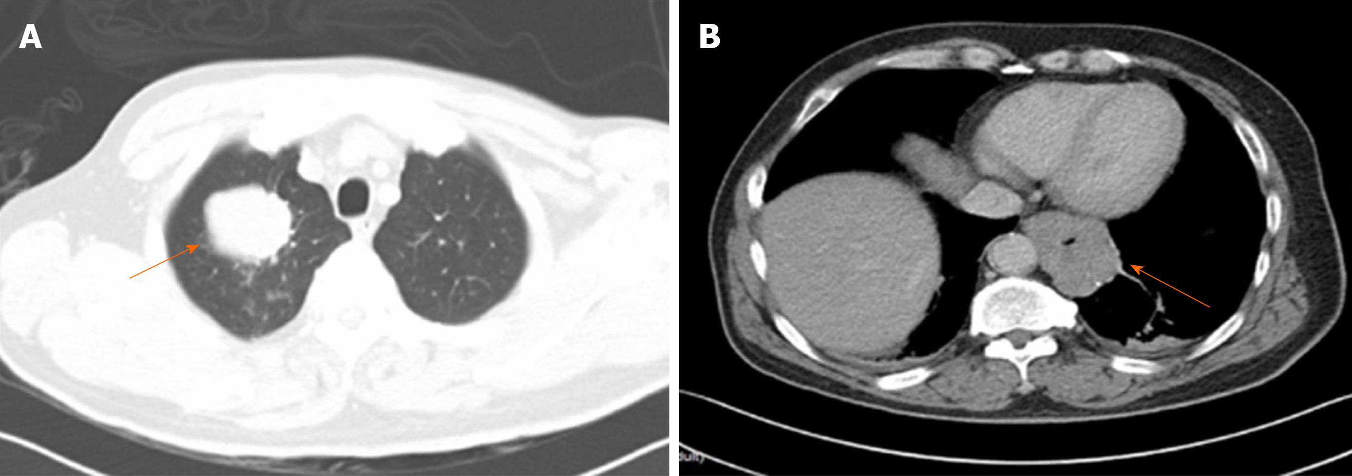
Figure 2 Preoperative chest computed tomography.
A: A right upper lobe mass; B: A lower esophageal and stomachus cardiacus mass.
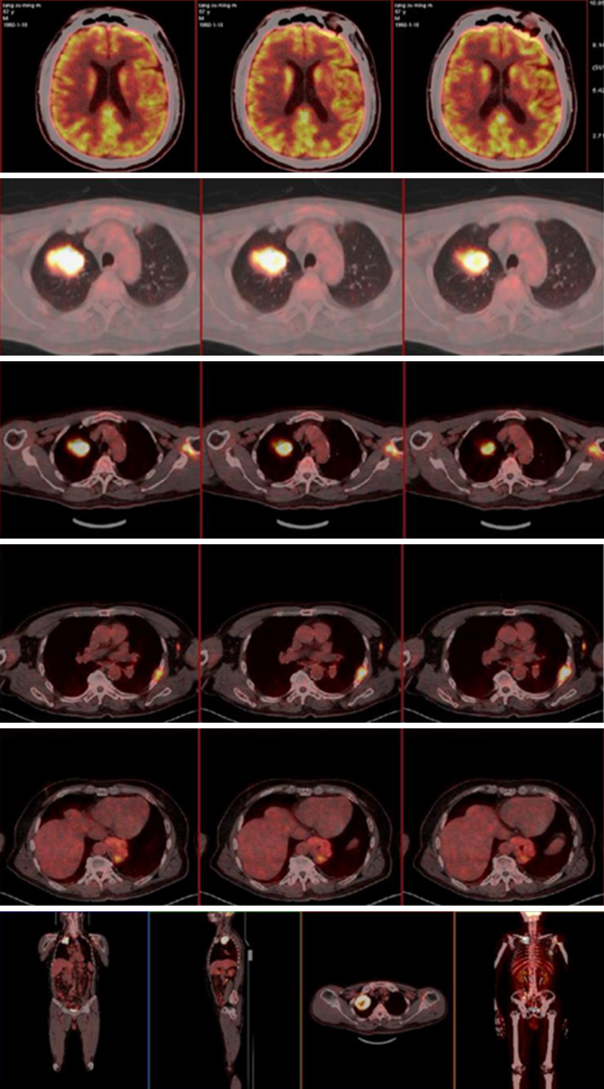
Figure 3 A preoperative 2-18F fluoro-2 deoxy-D-glucose positron emission tomography-computed tomography scan showed multiple malignant lesions throughout the whole body.
This led to a strong suspicion of gastroesophageal junction and/or right lung malignancies with multiple metastases.
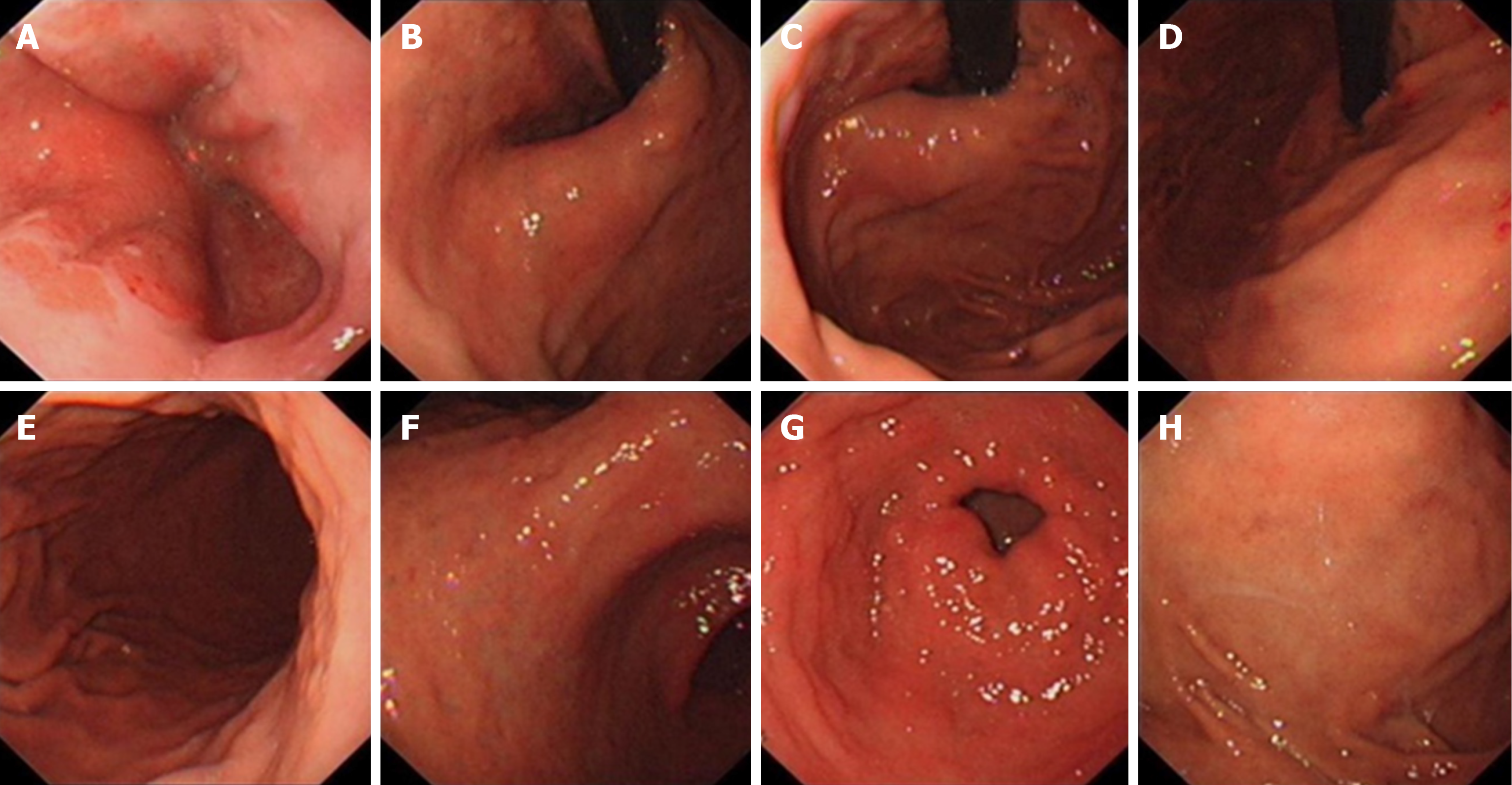
Figure 4 Preoperative gastroscopy did not find any space-occupying lesions.
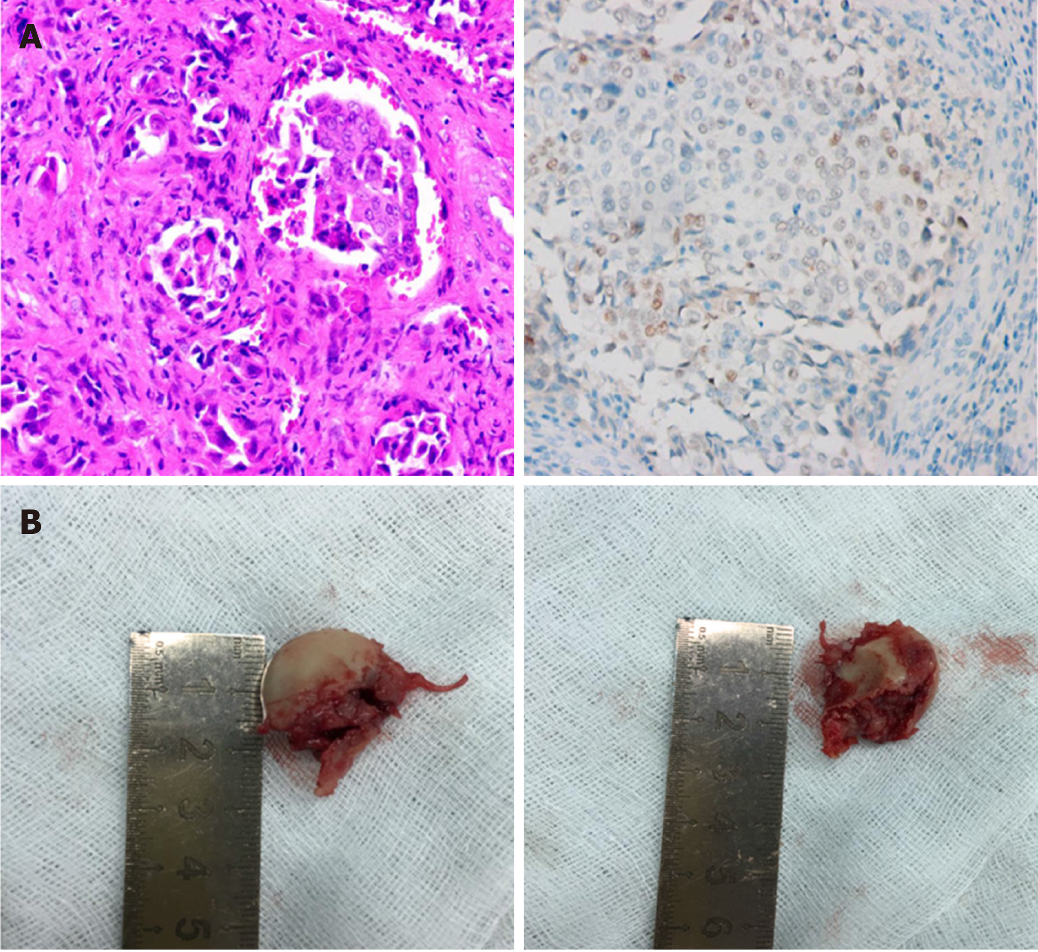
Figure 5 Intraoperative photographs and postoperative pathological results.
A: Pathological biopsy confirmed scaphoid bone metastasis of poorly differentiated adenocarcinoma. Immunohistochemistry showed CDX2 positivity (200×); B: The scaphoid bone was completely removed.
- Citation: Zhang YJ, Wang YY, Yang Q, Li JB. Scaphoid metastasis as the first sign of occult gastroesophageal junction cancer: A case report. World J Clin Cases 2020; 8(7): 1287-1294
- URL: https://www.wjgnet.com/2307-8960/full/v8/i7/1287.htm
- DOI: https://dx.doi.org/10.12998/wjcc.v8.i7.1287


