Copyright
©The Author(s) 2020.
World J Clin Cases. Apr 6, 2020; 8(7): 1278-1286
Published online Apr 6, 2020. doi: 10.12998/wjcc.v8.i7.1278
Published online Apr 6, 2020. doi: 10.12998/wjcc.v8.i7.1278
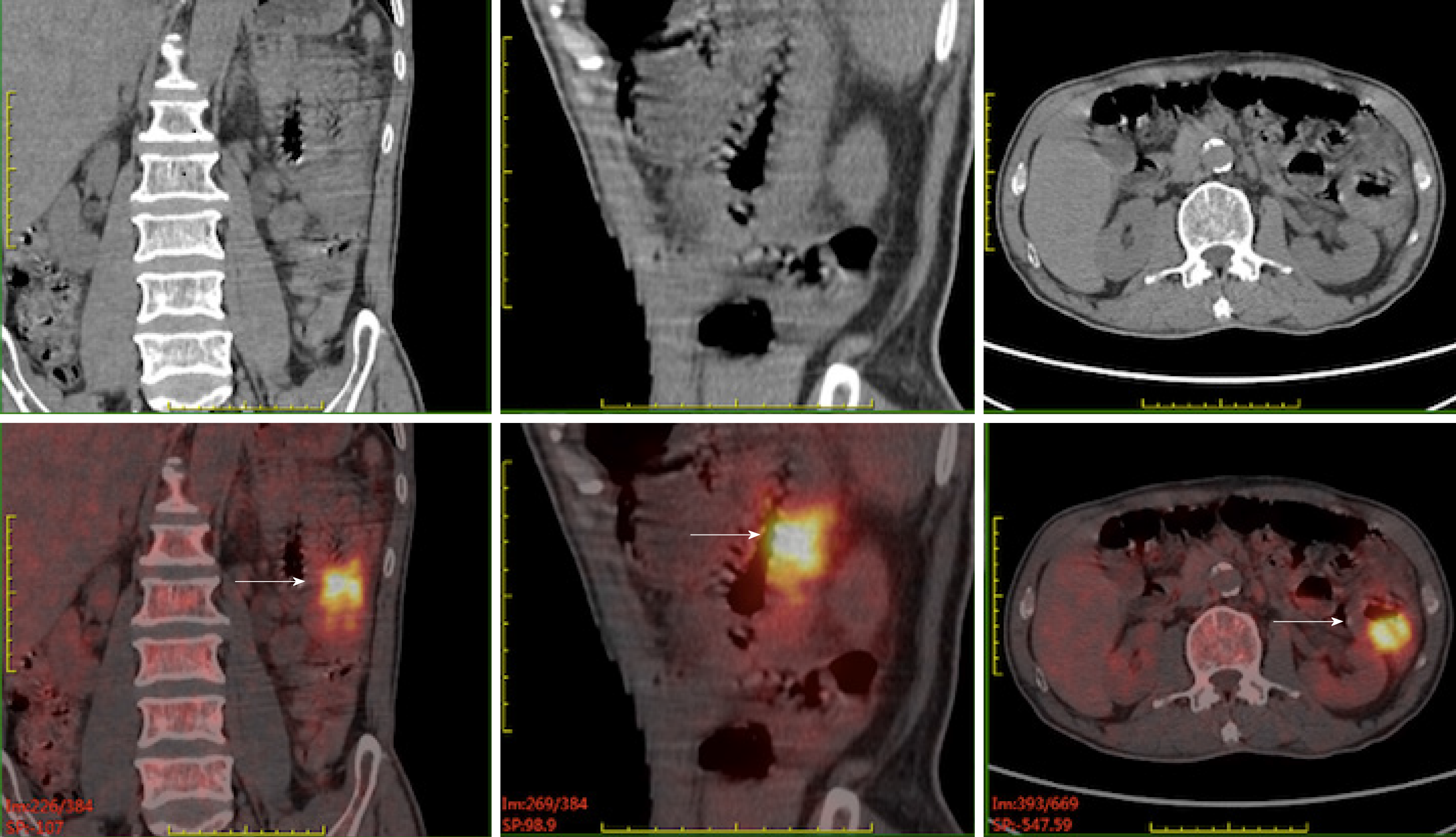
Figure 1 Pre-operative positron emission tomography/computed tomography of case one.
It revealed that the wall of the second group of the intestine was thickened with increased metabolism, and multiple enlarged and hypermetabolic lymph nodes were shown in the para-intestinal, mesenteric, and retroperitoneal regions.
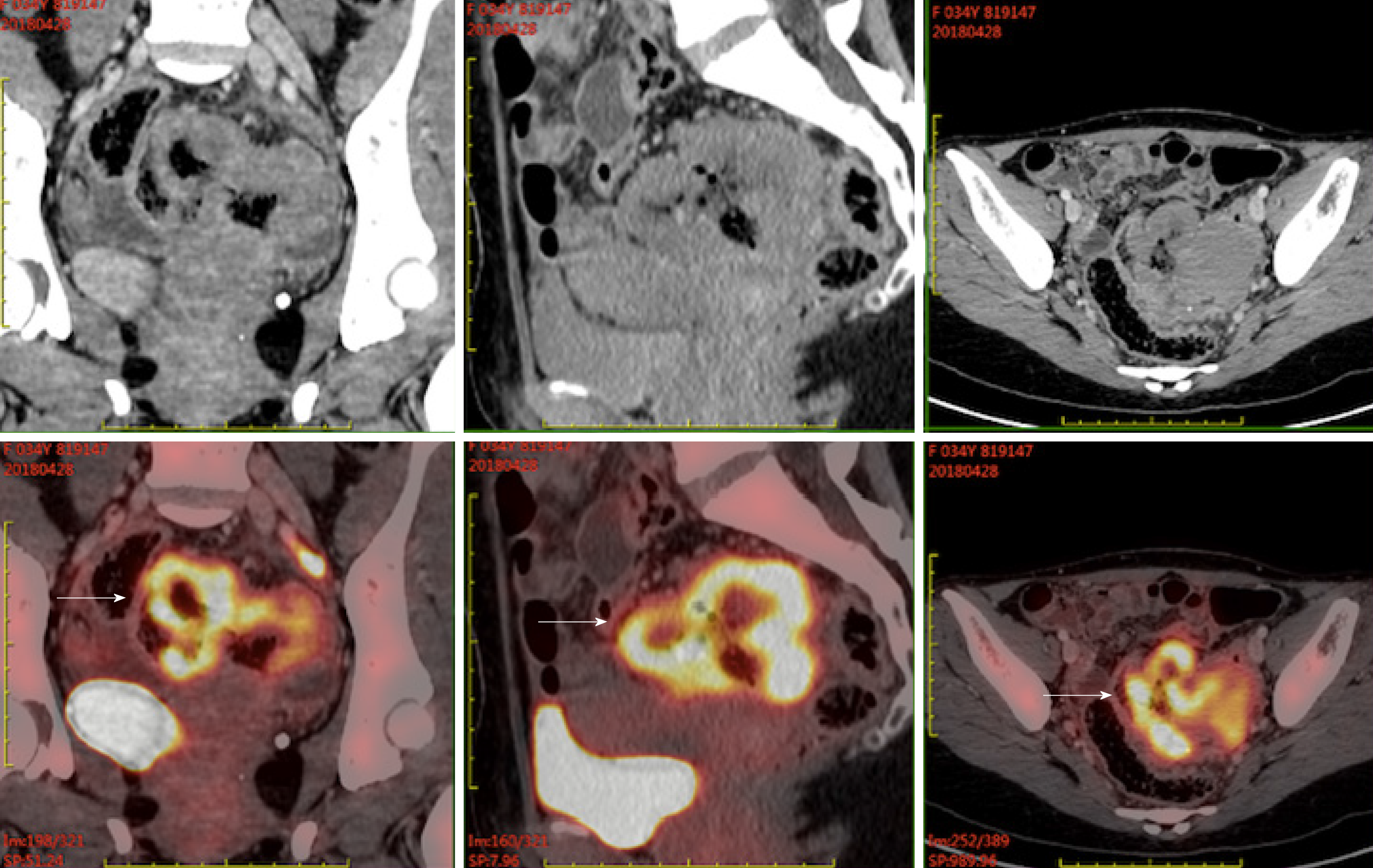
Figure 2 Pre-operative positron emission tomography/computed tomography of case two.
It demonstrated wall thickening of groups 1 and 4 of the small intestine and upper sigmoid colon with diffuse uptake. Small lymph nodes in the para-intestinal and mesenteric regions were also metabolically active.
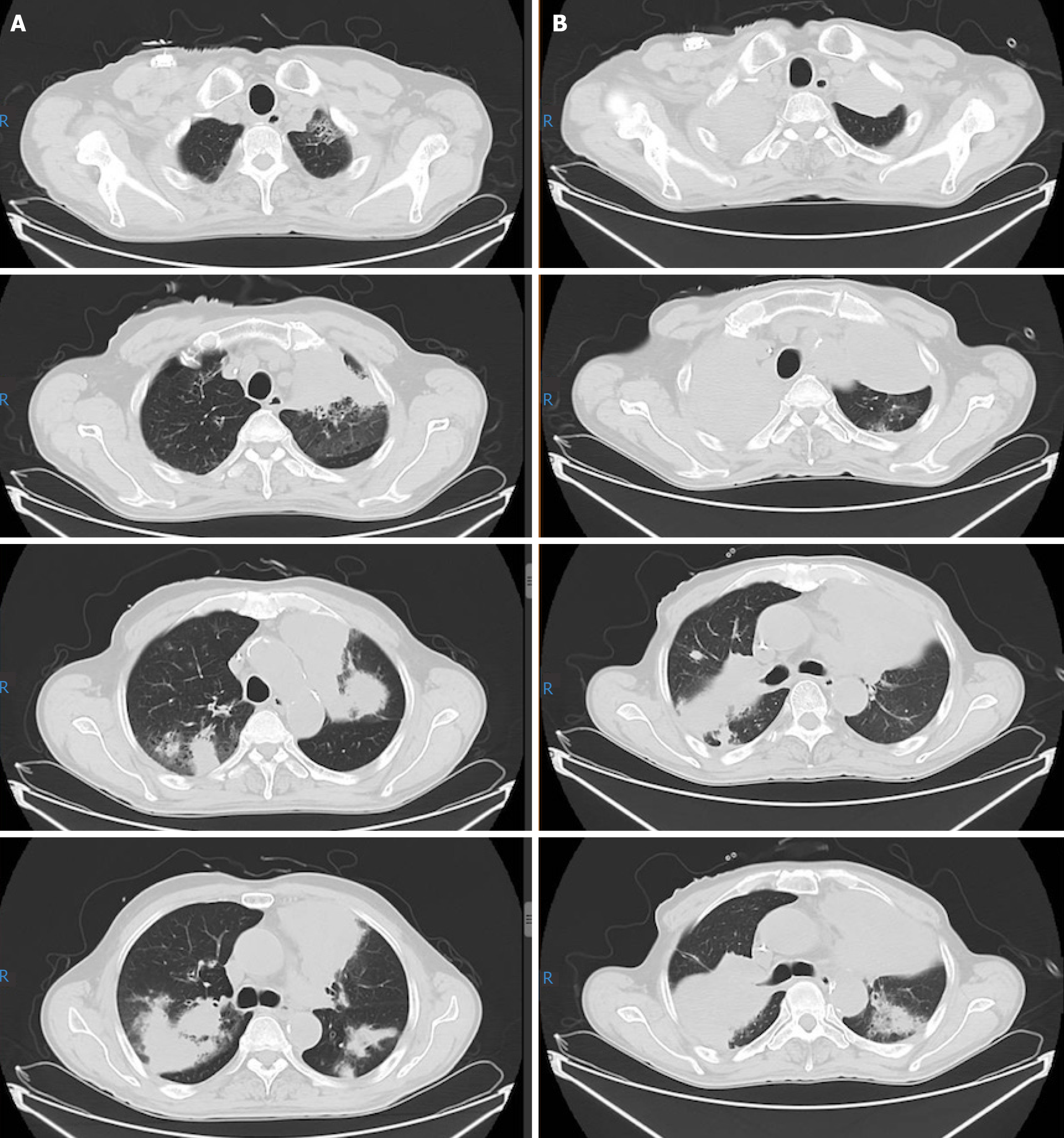
Figure 3 Thoracic computed tomography scan of case one taken at recurrence 11 mo following his diagnosis and re-evaluation after three cycles of rescue chemotherapy.
A: Computed tomography taken before rescue therapy. It revealed multiple lesions in the bilateral lungs; B: Computed tomography taken after rescue therapy. It showed that the shadow in bilateral lungs aggravated.
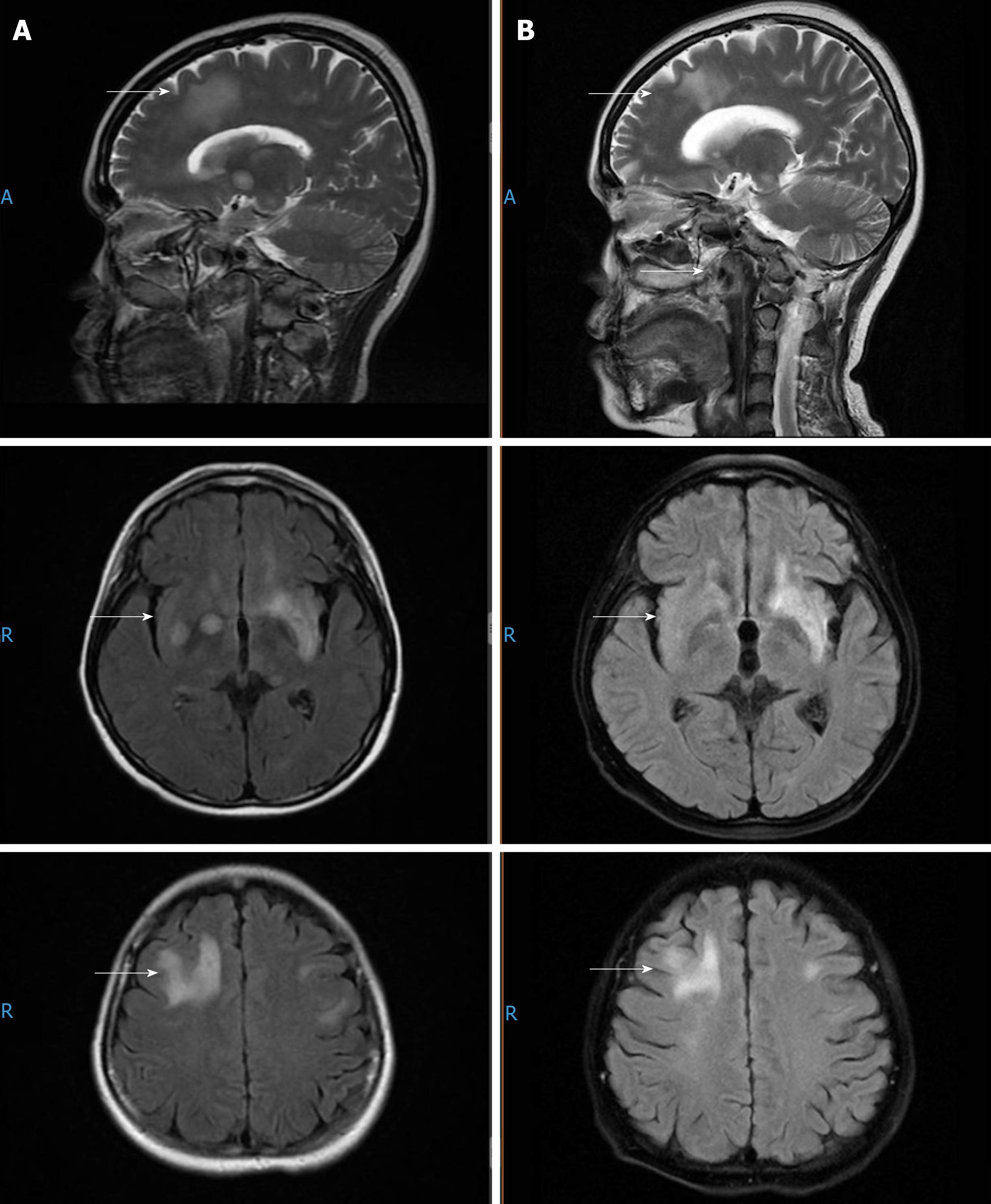
Figure 4 Head magnetic resonance imaging of case two taken at recurrence 6 mo following her diagnosis and re-evaluation after two cycles of rescue therapy.
A: Magnetic resonance taken before rescue therapy. It revealed multiple lesions in the bilateral frontotemporal lobes, semioval centers, basal ganglia, insular lobes, and left hippocampus and cerebral foot; B: Magnetic resonance taken after two cycles of rescue therapy. It showed the lesions in the bilateral brain shrank slightly.
- Citation: Liu TZ, Zheng YJ, Zhang ZW, Li SS, Chen JT, Peng AH, Huang RW. Chidamide based combination regimen for treatment of monomorphic epitheliotropic intestinal T cell lymphoma following radical operation: Two case reports. World J Clin Cases 2020; 8(7): 1278-1286
- URL: https://www.wjgnet.com/2307-8960/full/v8/i7/1278.htm
- DOI: https://dx.doi.org/10.12998/wjcc.v8.i7.1278


