Copyright
©The Author(s) 2020.
World J Clin Cases. Mar 6, 2020; 8(5): 995-1001
Published online Mar 6, 2020. doi: 10.12998/wjcc.v8.i5.995
Published online Mar 6, 2020. doi: 10.12998/wjcc.v8.i5.995
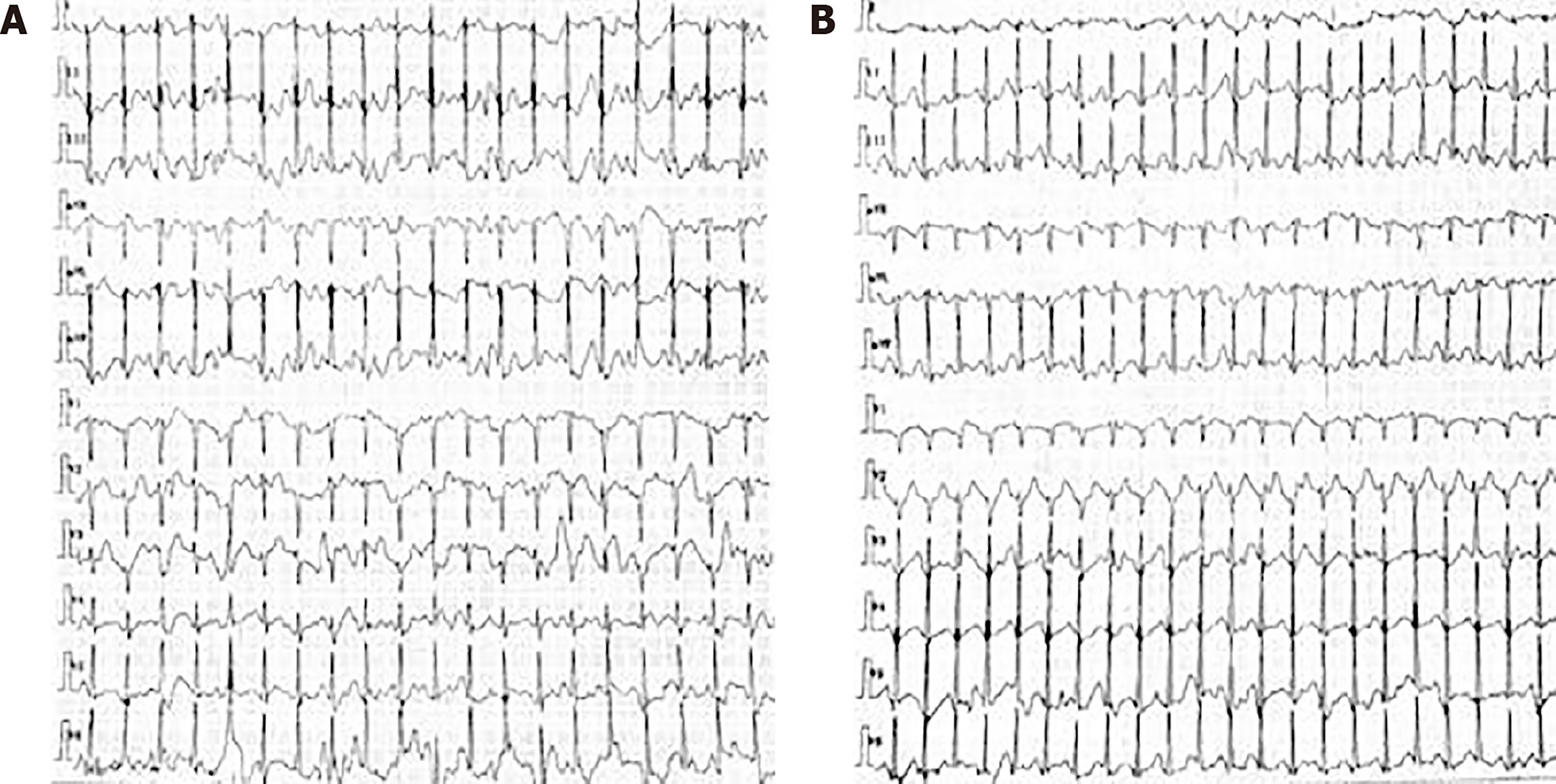
Figure 1 Holter electrocardiogram monitoring findings in the patient.
A: Holter electrocardiogram monitoring showed supraventricular tachycardia before treatment, and the heart rate was 158 bpm; B: Holter electrocardiogram monitoring showed supraventricular tachycardia when the patient presented with a loss of consciousness, and the heart rate was 184 bpm.
Figure 2 Myopathological findings in our patient.
A: Hematoxylin and eosin staining. Muscular fibers were mild variable in size and some muscular fibers were moderated to small vacuoles; B: Modified Gomori trichrome staining showed atypical ragged red fibers; C: Oil red O staining showed predominant lipid accumulation mainly in type I fibers.
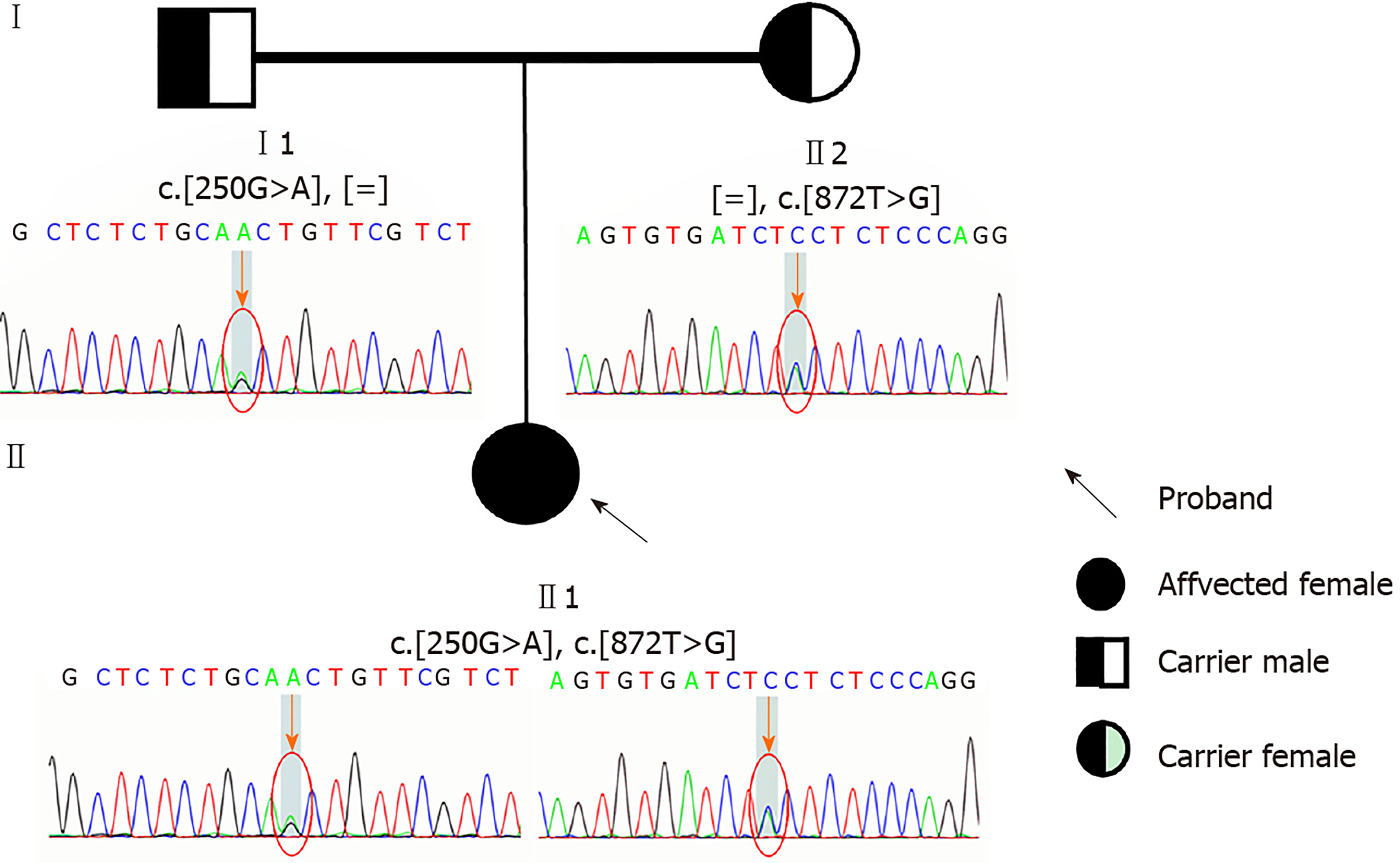
Figure 3 Result of gene sequencing about the patient and her parents.
Electropherogram of the heterozygous mutation c.250G>A (p.A84T) and c.872T>G (p.V291G) in the ETFDH gene on the patient; electropherogram of the heterozygous mutation c.250G>A (p.A84T) in the ETFDH gene on her father; and electropherogram of the heterozygous mutation c.872T>G (p.V291G) in the ETFDH gene on her mother are shown (arrows and circles indicate mutation site).
- Citation: Pan XQ, Chang XL, Zhang W, Meng HX, Zhang J, Shi JY, Guo JH. Late-onset multiple acyl-CoA dehydrogenase deficiency with cardiac syncope: A case report. World J Clin Cases 2020; 8(5): 995-1001
- URL: https://www.wjgnet.com/2307-8960/full/v8/i5/995.htm
- DOI: https://dx.doi.org/10.12998/wjcc.v8.i5.995


