Copyright
©The Author(s) 2020.
World J Clin Cases. Mar 6, 2020; 8(5): 986-994
Published online Mar 6, 2020. doi: 10.12998/wjcc.v8.i5.986
Published online Mar 6, 2020. doi: 10.12998/wjcc.v8.i5.986
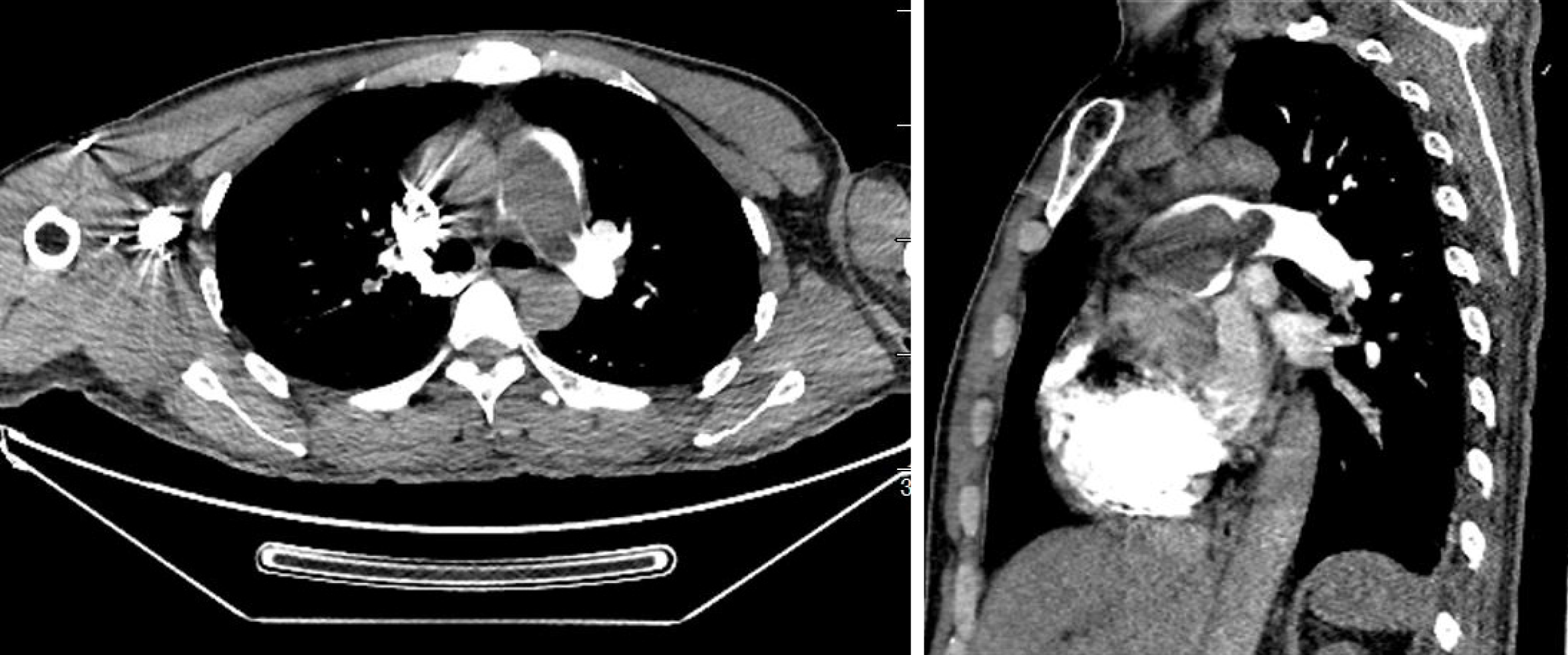
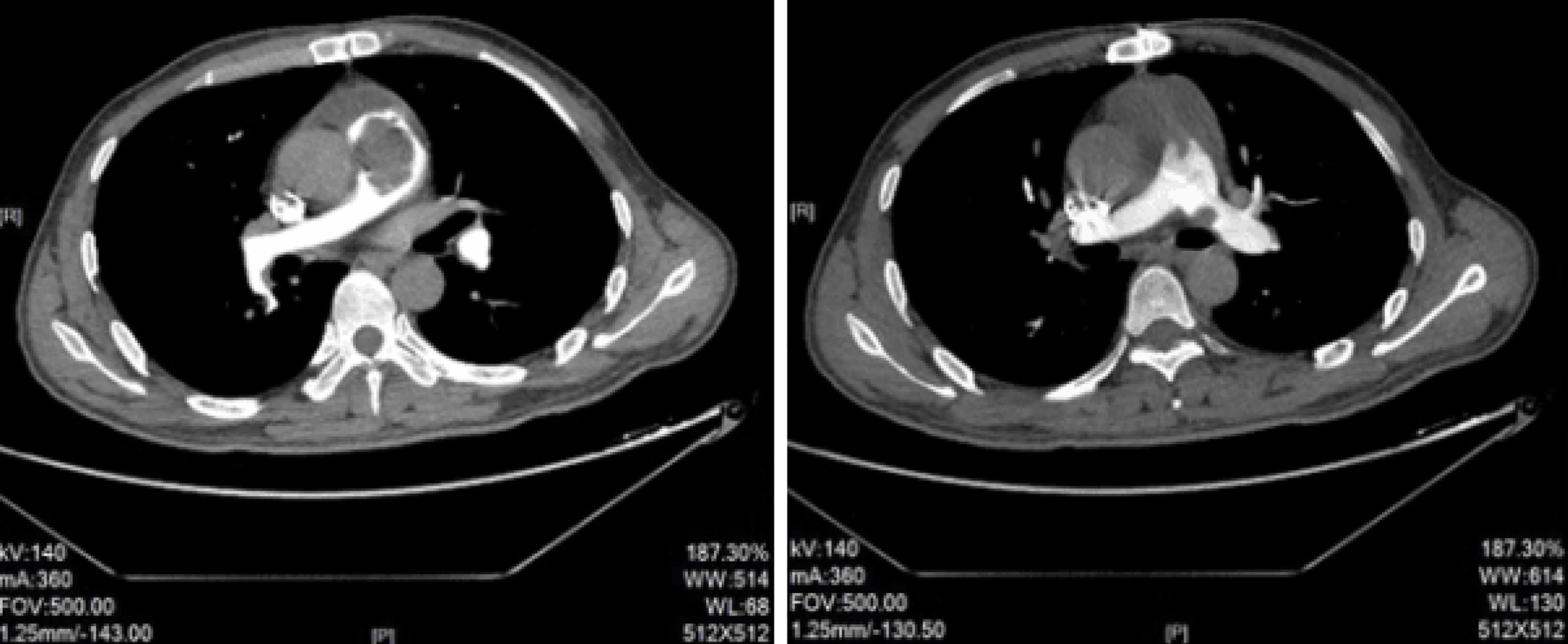
Figure 1 Preoperative computed tomography pulmonary angiography showed a significant filling defect in the pulmonary artery.
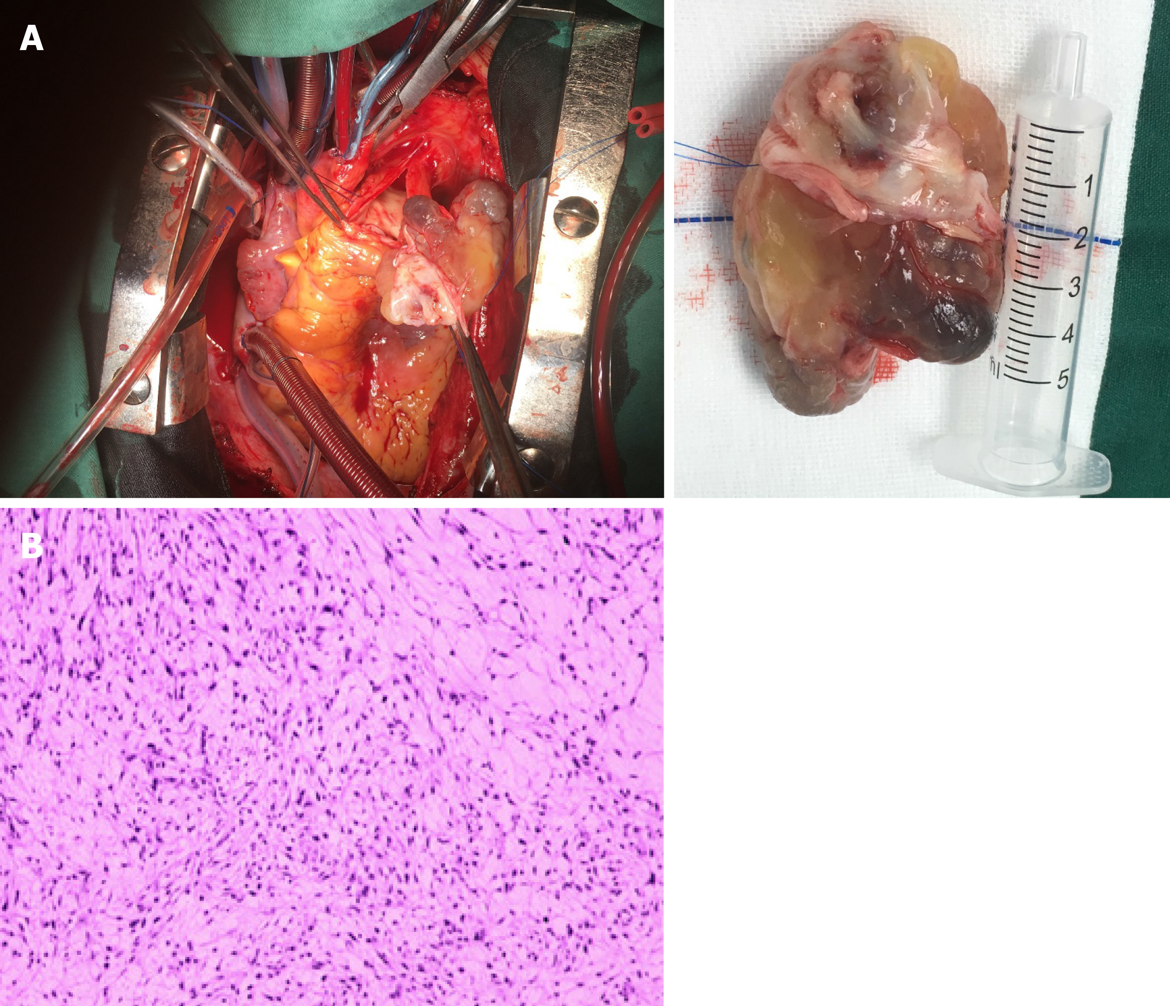
Figure 2 Intimal sarcoma of the pulmonary artery.
A: A giant pulmonary artery tumor was removed during surgery; B: Postoperative pathology showed intimal sarcoma of the pulmonary artery.
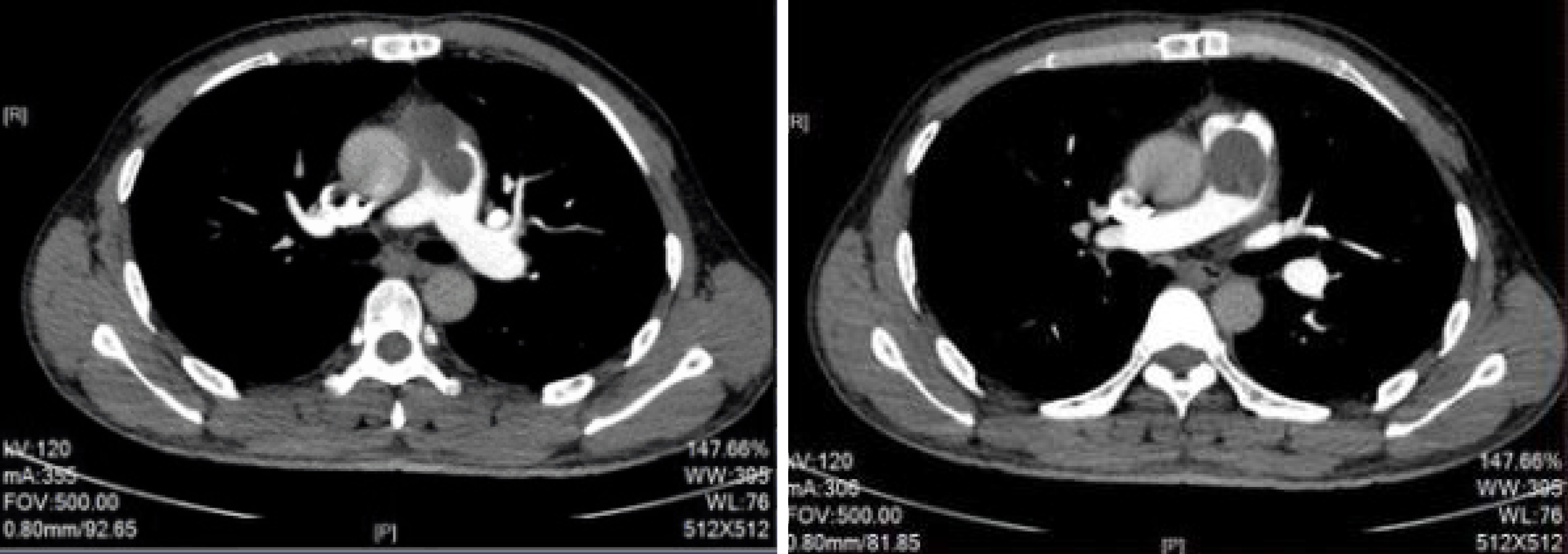
Figure 3 Computed tomography pulmonary angiography of the pulmonary artery at 3 mo post-operation showed relapse of the pulmonary artery sarcoma.
Figure 4 Computed tomography pulmonary angiography of the pulmonary artery after 2 mo of apatinib administration showed improved clinical conditions.
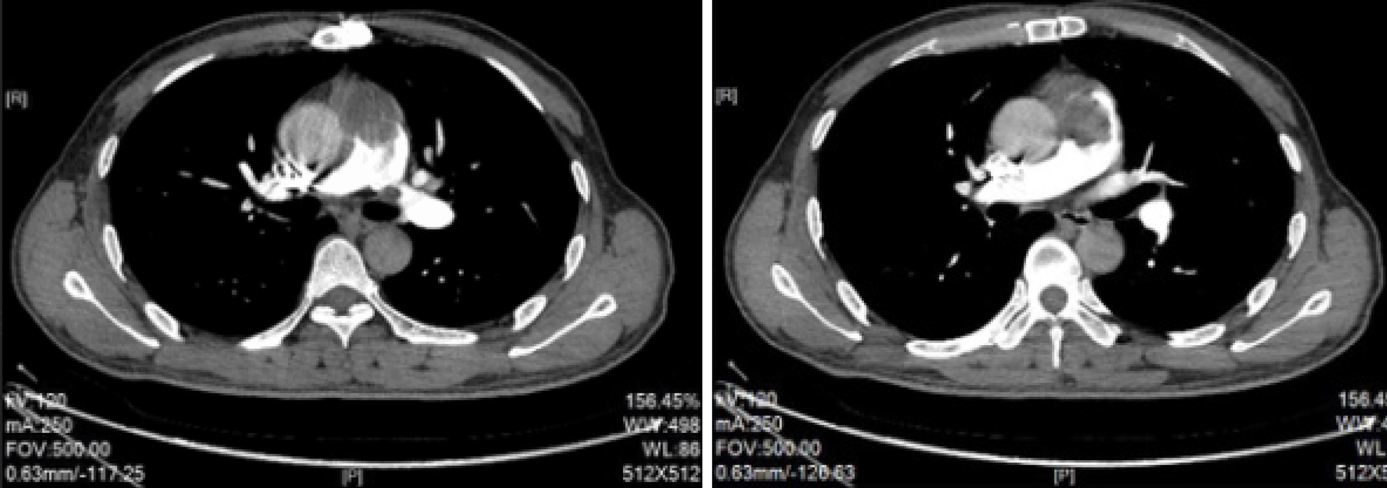
Figure 5 Computed tomography pulmonary angiography of the pulmonary artery after 4 mo of apatinib administration showed disease progression.
- Citation: Lu P, Yin BB. Misdiagnosis of primary intimal sarcoma of the pulmonary artery as chronic pulmonary embolism: A case report. World J Clin Cases 2020; 8(5): 986-994
- URL: https://www.wjgnet.com/2307-8960/full/v8/i5/986.htm
- DOI: https://dx.doi.org/10.12998/wjcc.v8.i5.986


