Copyright
©The Author(s) 2020.
World J Clin Cases. Mar 6, 2020; 8(5): 946-953
Published online Mar 6, 2020. doi: 10.12998/wjcc.v8.i5.946
Published online Mar 6, 2020. doi: 10.12998/wjcc.v8.i5.946
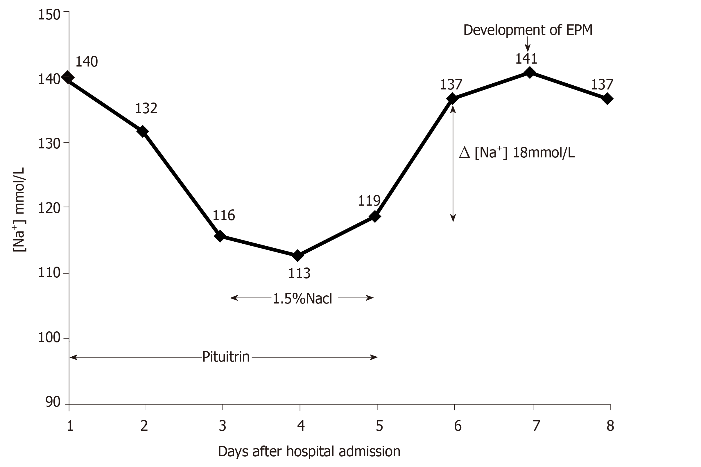
Figure 1 Serum sodium levels of the patient after admission.
The serum sodium gradually declined after the use of pituitrin. Hypertonic saline was administered on day 3 until day 5. Pituitrin therapy was stopped on day 5, then a sharp increase in serum sodium level was detected on day 6, and neurological symptoms appeared on the seventh day after admission.
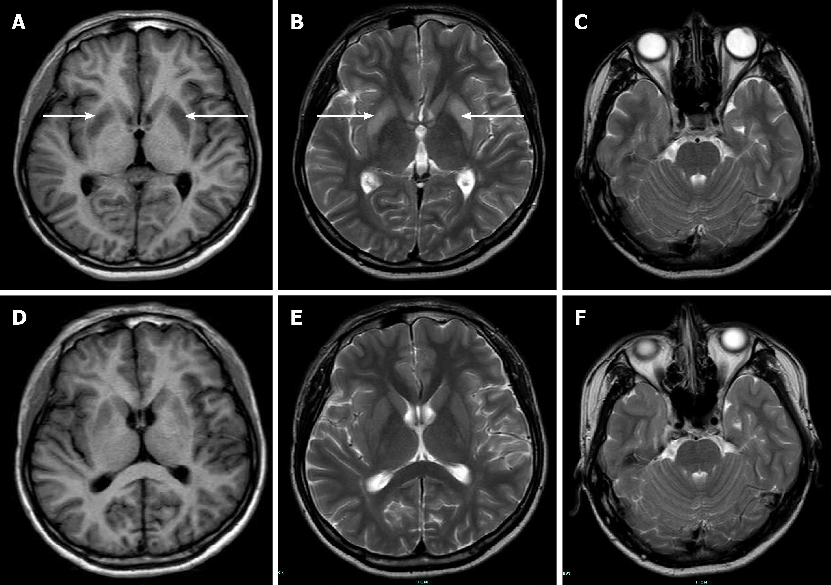
Figure 2 Brain magnetic resonance imaging during hospitalization.
A: Initial brain magnetic resonance imaging (MRI) obtained 3 d after neurological deterioration displayed low signal intensity in the bilateral symmetric basal ganglia (the white arrows) on the axial T1-weighted MRI; B: High signal intensity (the white arrows) on the axial T2-weighted images; C: No lesion in the pons; D-F: Follow-up brain MRI performed one month after treatment. E: Compared to the previous brain MRI, a markedly decreased signal was observed on the T2-weighted image, indicating a significant improvement.
- Citation: Fang LJ, Xu MW, Zhou JY, Pan ZJ. Extrapontine myelinolysis caused by rapid correction of pituitrin-induced severe hyponatremia: A case report. World J Clin Cases 2020; 8(5): 946-953
- URL: https://www.wjgnet.com/2307-8960/full/v8/i5/946.htm
- DOI: https://dx.doi.org/10.12998/wjcc.v8.i5.946


