Copyright
©The Author(s) 2020.
World J Clin Cases. Feb 6, 2020; 8(3): 645-651
Published online Feb 6, 2020. doi: 10.12998/wjcc.v8.i3.645
Published online Feb 6, 2020. doi: 10.12998/wjcc.v8.i3.645
Figure 1 Enhanced chest computed tomography.
Computed tomography shows local thickening of the esophageal wall with moderate enhancement.
Figure 2 Upper gastrointestinal endoscopy.
A 1.5 cm plate-shaped ulcerated hyperplastic lesion is seen in the middle esophagus, with a central depression, erosion, and slight bleeding.
Figure 3 Endoscopic ultrasonography.
A homogeneous hypoechoic lesion protrudes into the esophageal lumen, with disappearance and fusion of the mucosal layers, and 17.5 mm × 21.3 mm enlarged lymph nodes adjacent to the esophageal lesion.
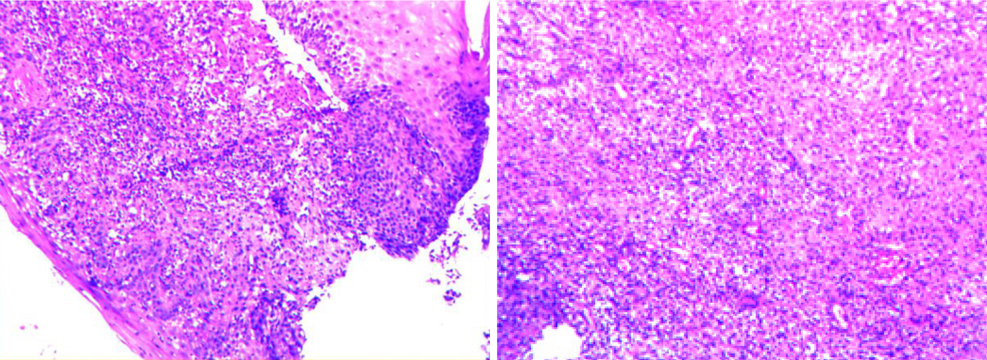
Figure 4 Pathologic biopsy of esophageal lesions.
Hematoxylin-eosin staining, × 200 shows epithelial detachment and interstitial granulation tissue hyperplasia, under inflammatory exudation.
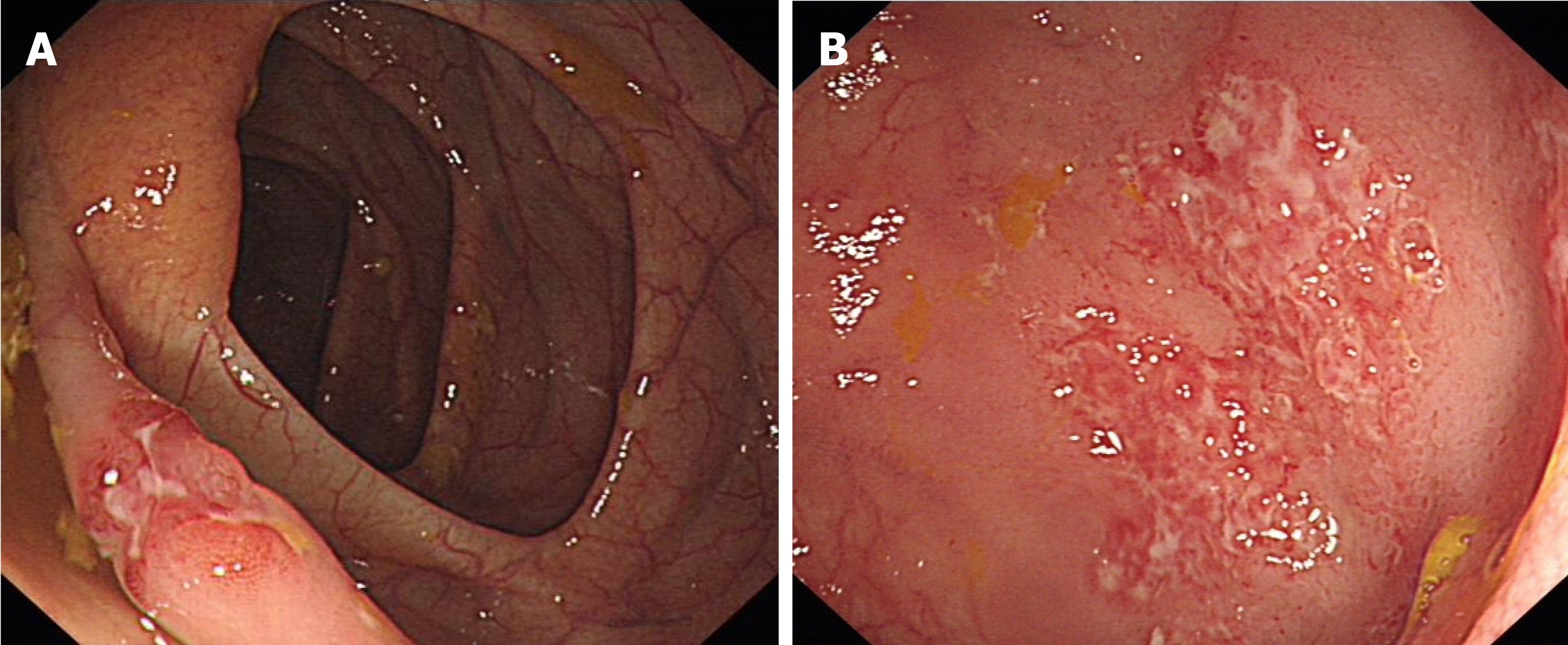
Figure 5 Colonoscopy.
A: An irregular ulcer with a rat-bite border in the terminal ileum; B: A shallow ulcer with irregular border in the ascending colon.
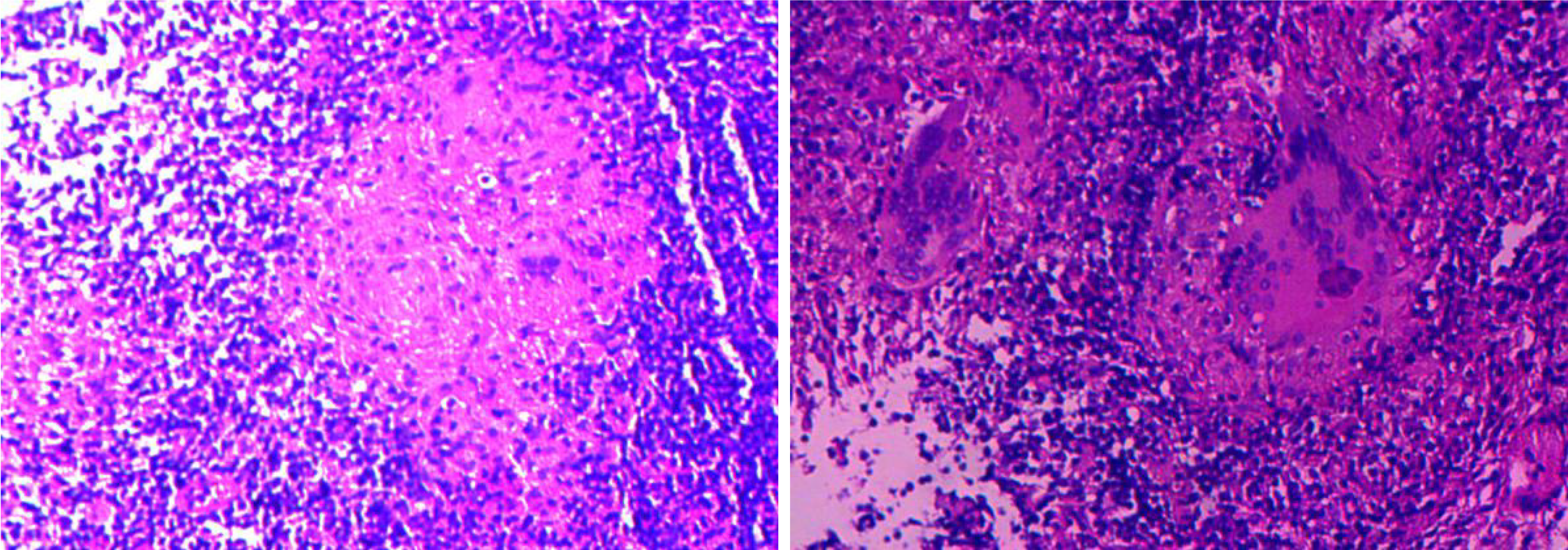
Figure 6 Pathologic biopsy of the terminal ileal lesion.
Hematoxylin-eosin staining × 200 reveals that the mucosa was in an acute inflammatory activity period of chronic inflammation, with decreasing or disappearing crypts in some areas, and some small focal granulomas, with caseous necrosis and surrounded by numerous lymphocytes.
Figure 7 Follow-up upper gastrointestinal endoscopy.
After 2 mo of anti-tuberculosis therapy, the endoscopy shows ulcer healing with scar tissue.
- Citation: Mao L, Zhou XT, Li JP, Li J, Wang F, Ma HM, Su XL, Wang X. Esophageal tuberculosis complicated with intestinal tuberculosis: A case report. World J Clin Cases 2020; 8(3): 645-651
- URL: https://www.wjgnet.com/2307-8960/full/v8/i3/645.htm
- DOI: https://dx.doi.org/10.12998/wjcc.v8.i3.645


