Copyright
©The Author(s) 2020.
World J Clin Cases. Feb 6, 2020; 8(3): 638-644
Published online Feb 6, 2020. doi: 10.12998/wjcc.v8.i3.638
Published online Feb 6, 2020. doi: 10.12998/wjcc.v8.i3.638
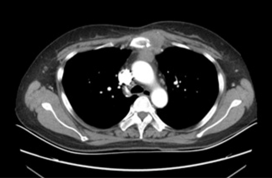
Figure 1 Chest contrast enhanced computed tomography scan.
Patchy low-density shadows were seen in the upper sternum, and soft tissue mass formation was observed around the upper sternum, with a size of 3.9 cm × 2.8 cm and slight enhancement.
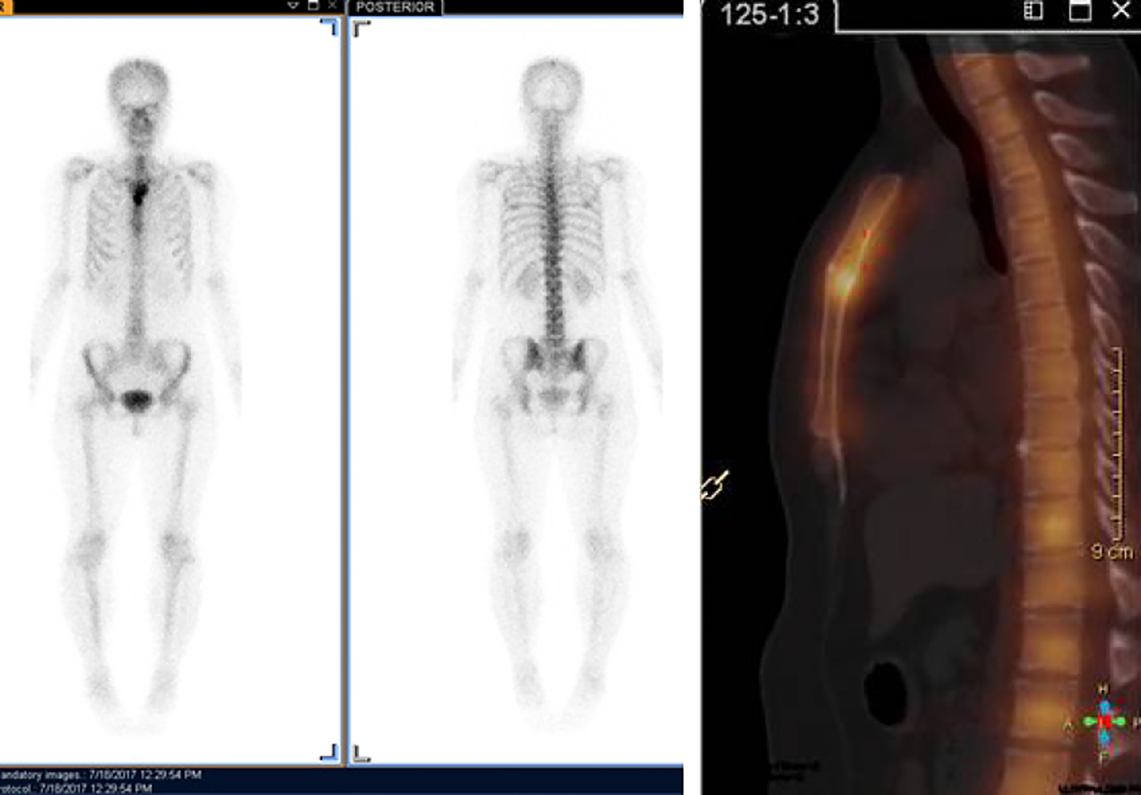
Figure 2 Bone scintigraphy scan with Tc99-MDP.
Apparent fixation of the radiopharmaceuticals in the manubrium sternum and upper segment of the body of the sternum. No involvement of other sites of the skeleton was found.
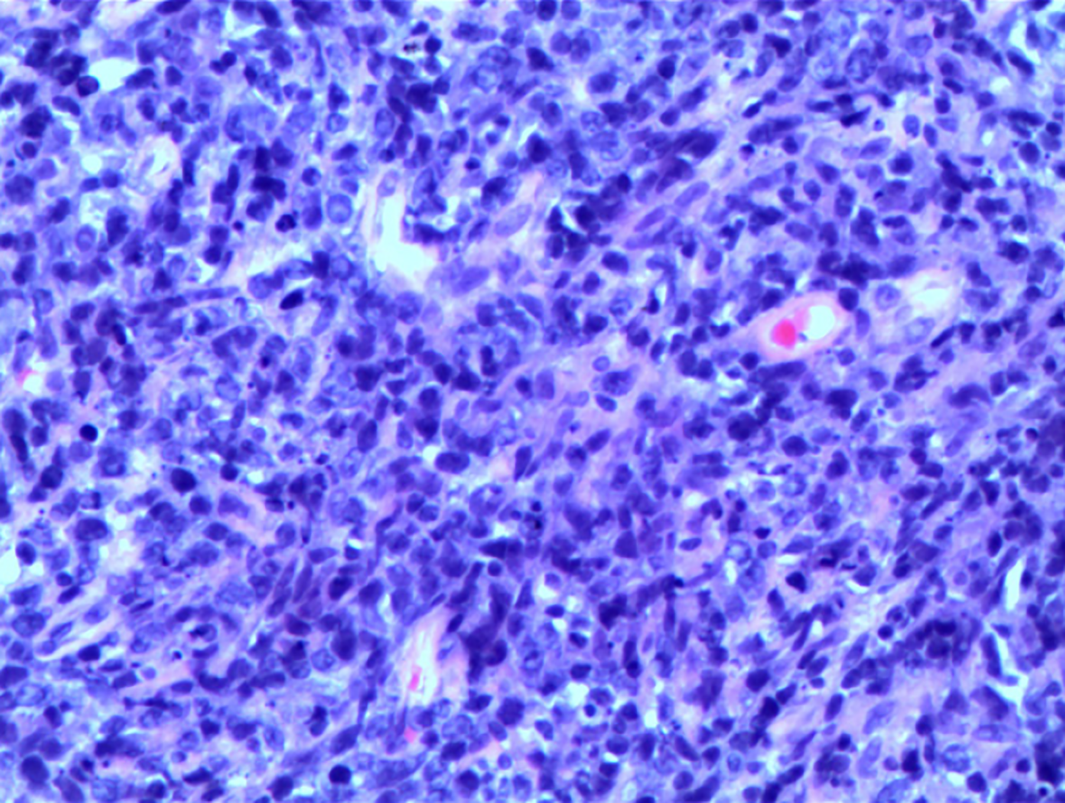
Figure 3 Hematoxylin and eosin staining (original magnification 200 ×).
Lymphocytes, plasma cells and a few large heteromorphic cells (Suspected Reed Sternberg cells).
Figure 4 Immunohistochemical staining (original magnification 100 ×).
A: CD 30(+); B: PAX-5(+); C: Ki-67(60%+).
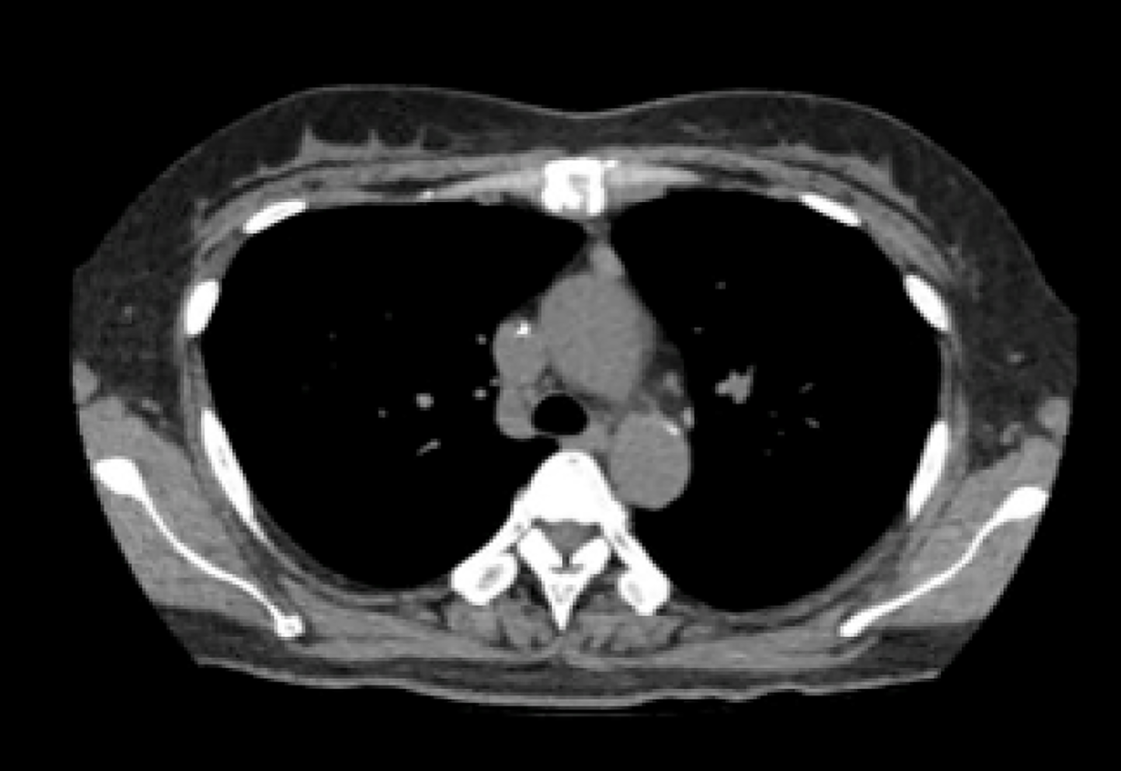
Figure 5 Chest computed tomography shows that the mass around the upper sternum was reduced obviously.
- Citation: Yin YY, Zhao N, Yang B, Xin H. Sternal Hodgkin’s lymphoma: A case report and review of literature. World J Clin Cases 2020; 8(3): 638-644
- URL: https://www.wjgnet.com/2307-8960/full/v8/i3/638.htm
- DOI: https://dx.doi.org/10.12998/wjcc.v8.i3.638


