Copyright
©The Author(s) 2020.
World J Clin Cases. Feb 6, 2020; 8(3): 624-629
Published online Feb 6, 2020. doi: 10.12998/wjcc.v8.i3.624
Published online Feb 6, 2020. doi: 10.12998/wjcc.v8.i3.624
Figure 1 Initial aspects of the gingiva.
A: A gingival mass extending from canine to canine on the lingual surfaces of the mandible; B: A moderate gingival overgrowth presenting on facial aspects of the lower anterior region; C: An interdental papilla enlargement in the upper anterior region.
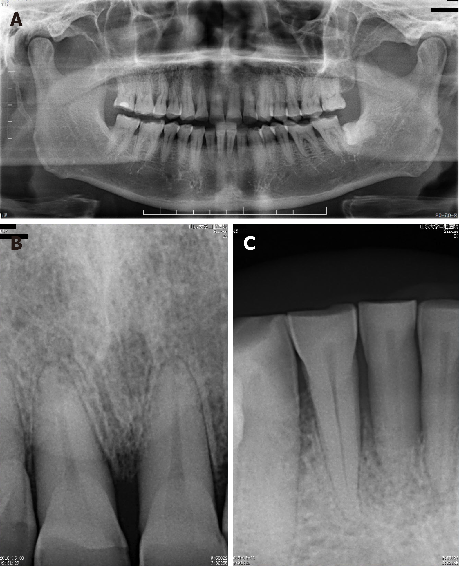
Figure 2 Radiographic aspects.
A: Moderate horizontal bone resorption in the upper and lower alveolar bone; B: Mild horizontal bone resorption in the upper front teeth; C: Severe horizontal bone resorption in teeth 31 and 41.
Figure 3 Histopathological findings.
A: Many blood vessels in the connective tissue stroma (Hematoxylin and eosin staining; magnification, 40 ×); B: Stratified squamous surface epithelium (Hematoxylin and eosin staining; magnification, 100 ×); C: The connective tissue stroma was composed of loosely arranged collagen fiber bundles interspersed with moderate chronic inflammatory cell infiltration of lymphocytes and plasma cells and many blood vessels containing red blood cells (Hematoxylin and eosin staining; magnification, 100 ×).
Figure 4 Gingival aspects at two weeks after surgery.
A: Surgical site healed uneventfully with gingival color normalization and slight swelling; B and C: The interdental papilla overgrowth around the upper anterior teeth.
- Citation: Yu Q, Wang WX. Camrelizumab (SHR-1210) leading to reactive capillary hemangioma in the gingiva: A case report. World J Clin Cases 2020; 8(3): 624-629
- URL: https://www.wjgnet.com/2307-8960/full/v8/i3/624.htm
- DOI: https://dx.doi.org/10.12998/wjcc.v8.i3.624


