Copyright
©The Author(s) 2020.
World J Clin Cases. Feb 6, 2020; 8(3): 614-623
Published online Feb 6, 2020. doi: 10.12998/wjcc.v8.i3.614
Published online Feb 6, 2020. doi: 10.12998/wjcc.v8.i3.614
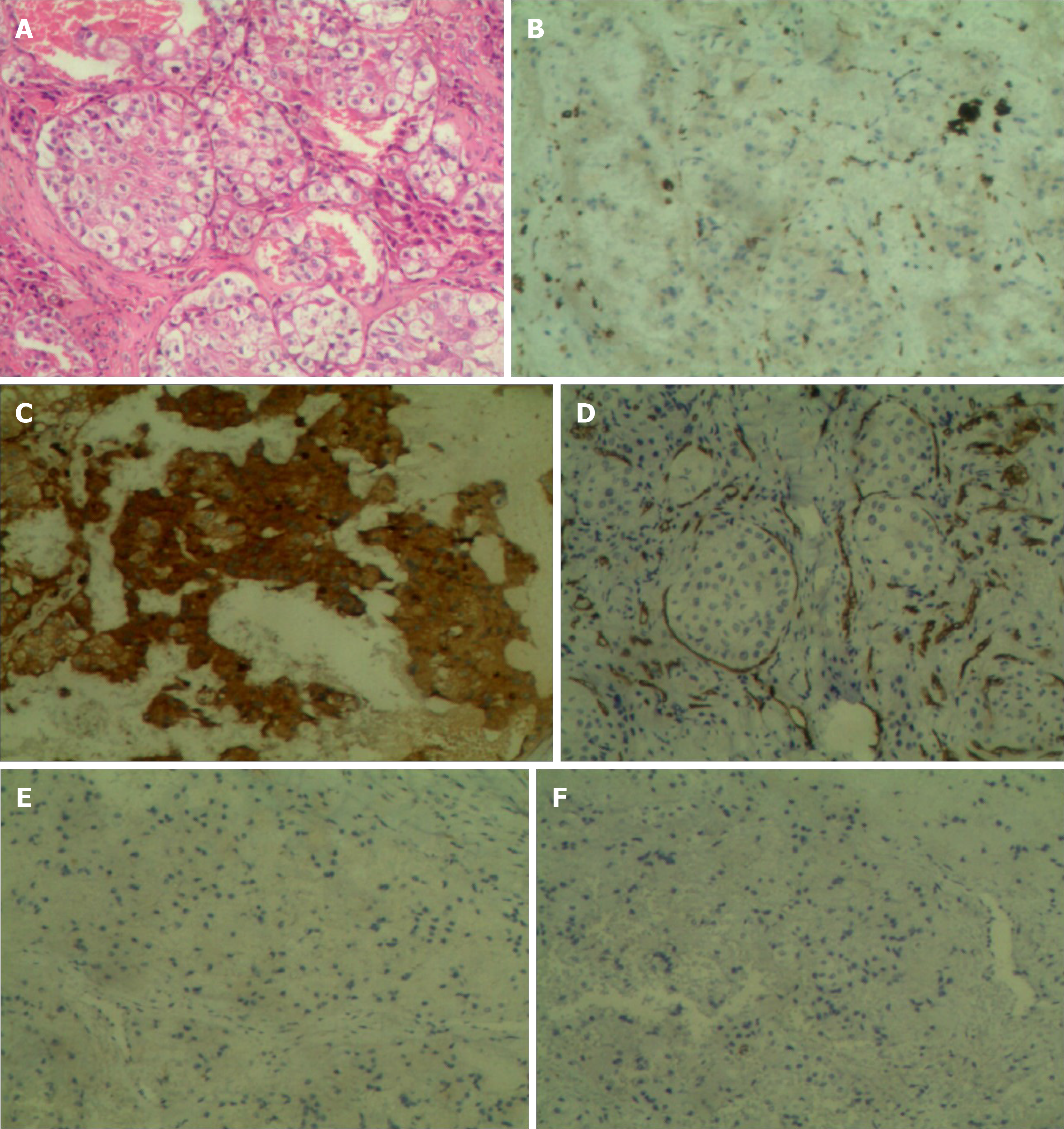
Figure 1 Histopathological findings of the lesions (100 ×).
A: Hematoxylin-eosin stains of the specimen; B: Immunohistochemistry showing positive staining for CD68; C: Immunohistochemistry showing positive staining for ACTT; D: Immunohistochemistry showing negative staining for CD34; E: Immunohistochemistry showing negative staining for S100; F: Immunohistochemistry showing negative staining for CD1a.
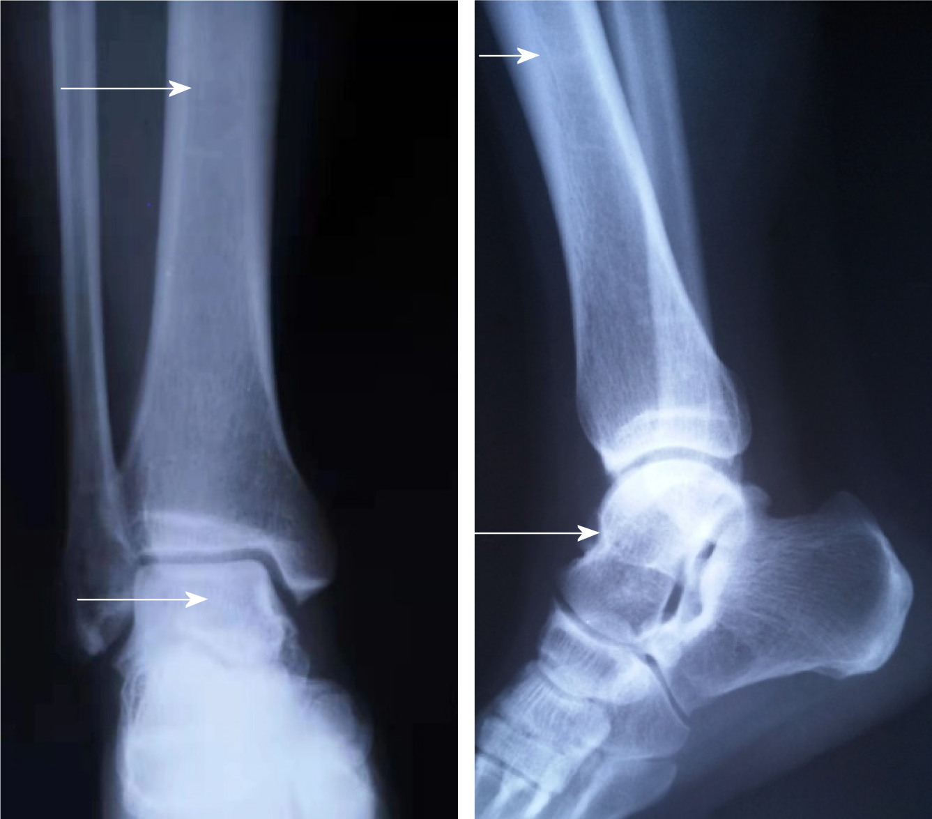
Figure 2 Plain X-ray film taken 5 years ago showed slight osteosclerosis in the tibia and the talus (white arrow).
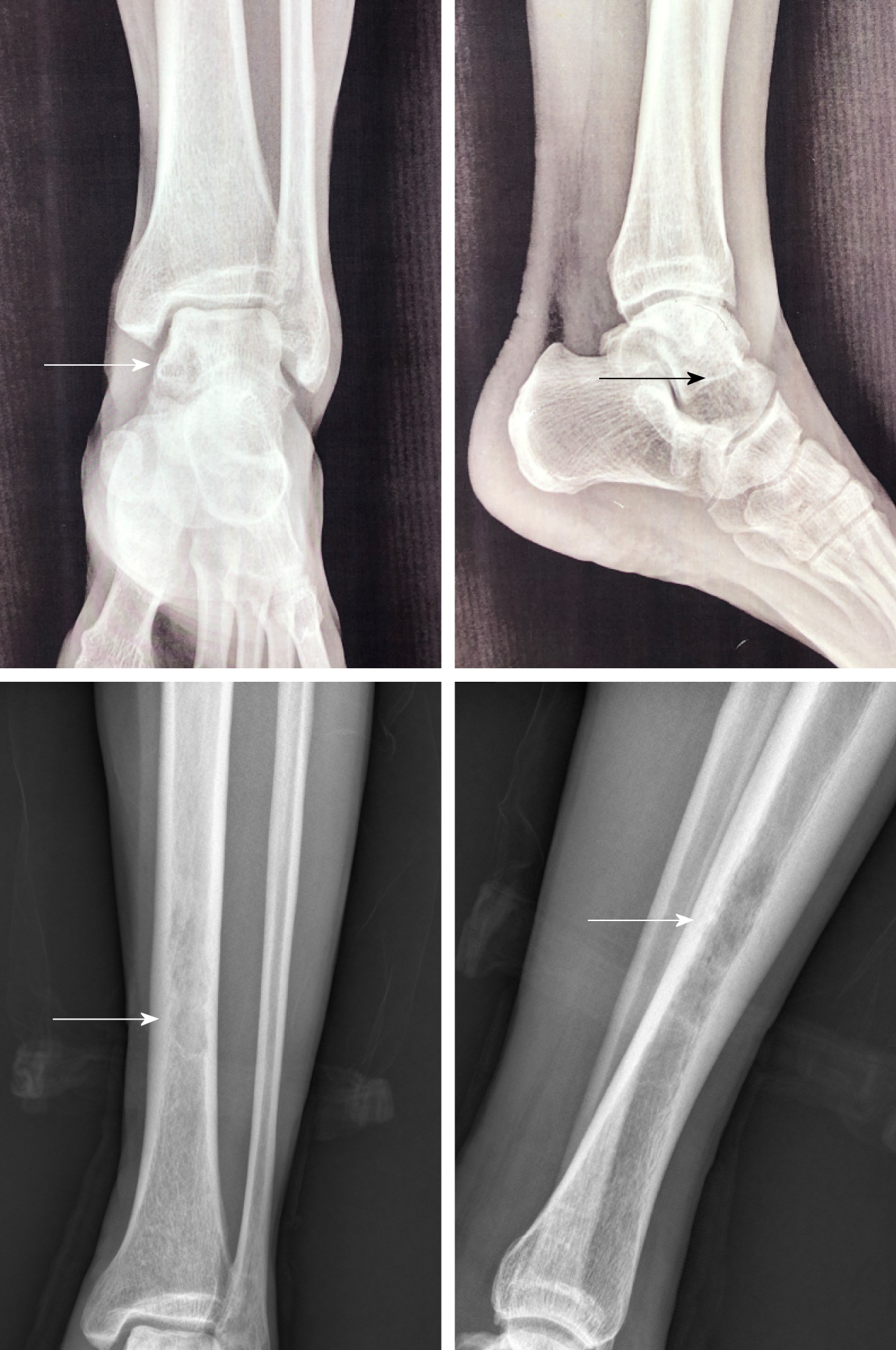
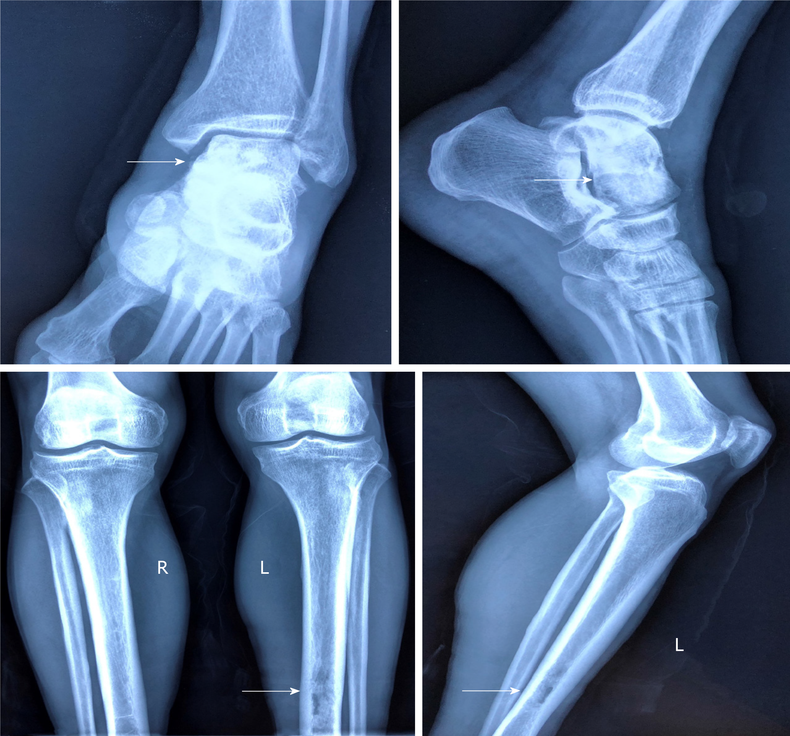
Figure 3 Plain anteroposterior radiography of the tibiofibula indicated osteosclerosis in the tibia and osteolytic lesions in the talus (arrow).
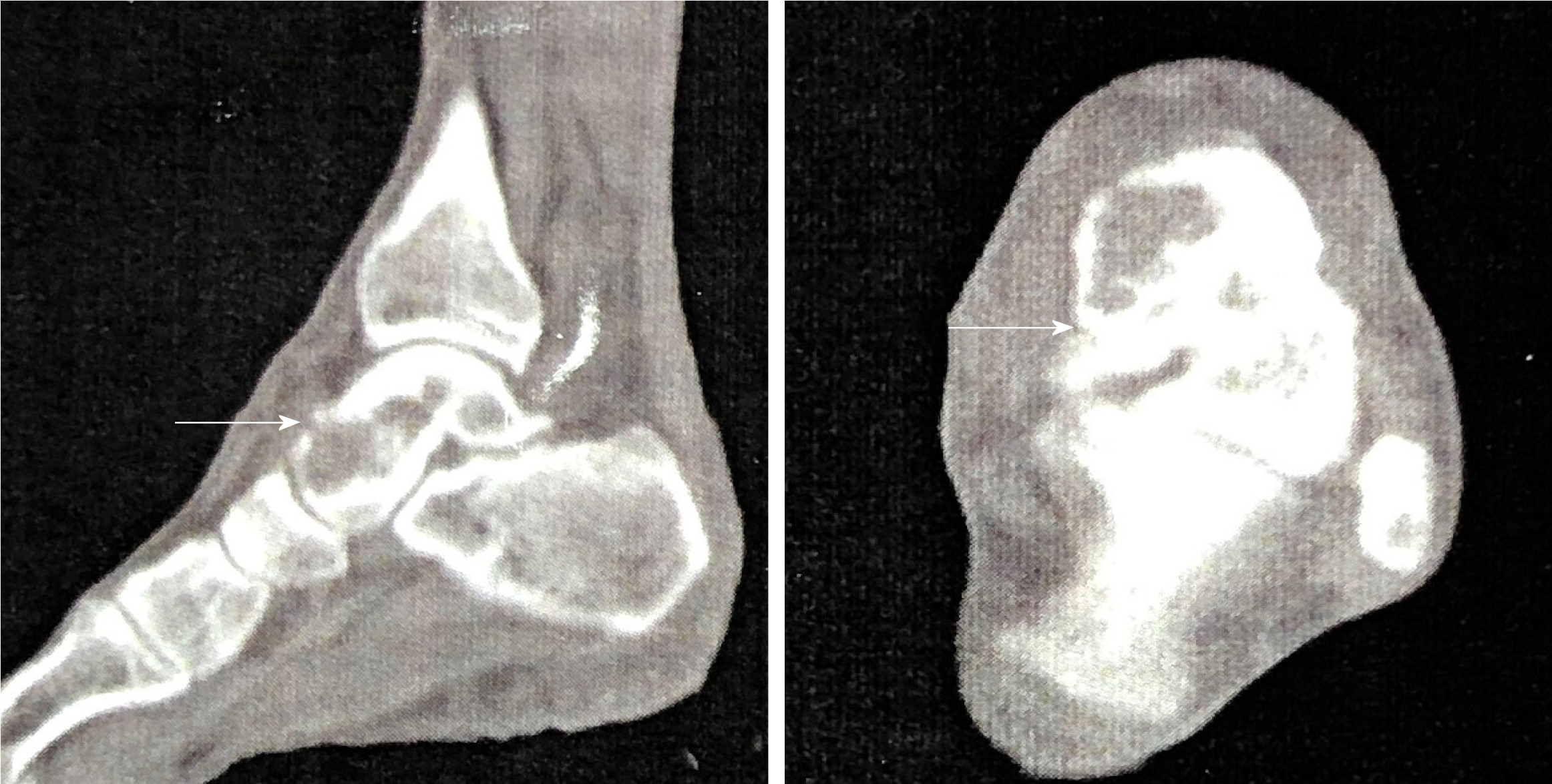
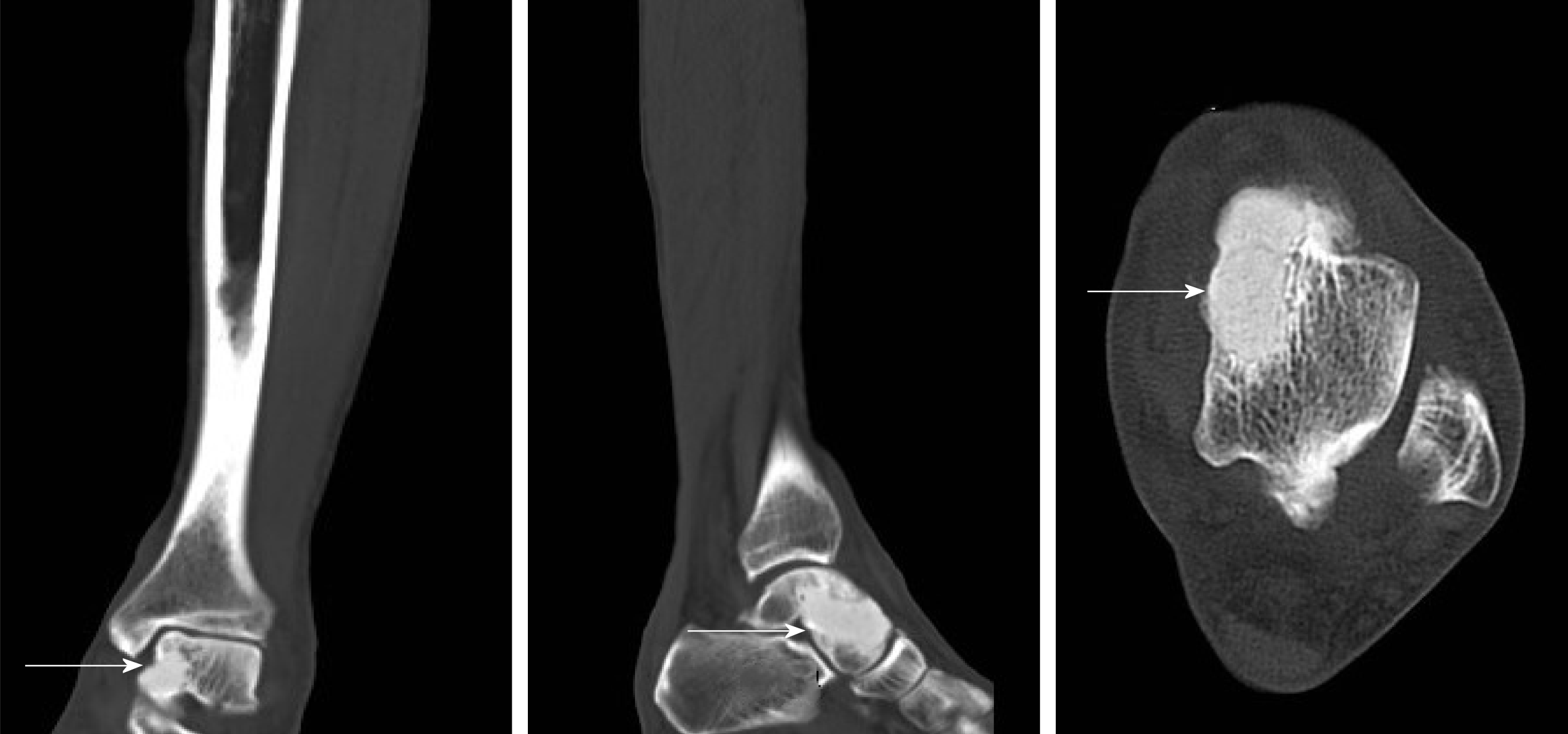
Figure 4 Computed tomography clearly suggested the left talus osteolytic destruction and cortical discontinuity of the anterior articular surface (white arrow).
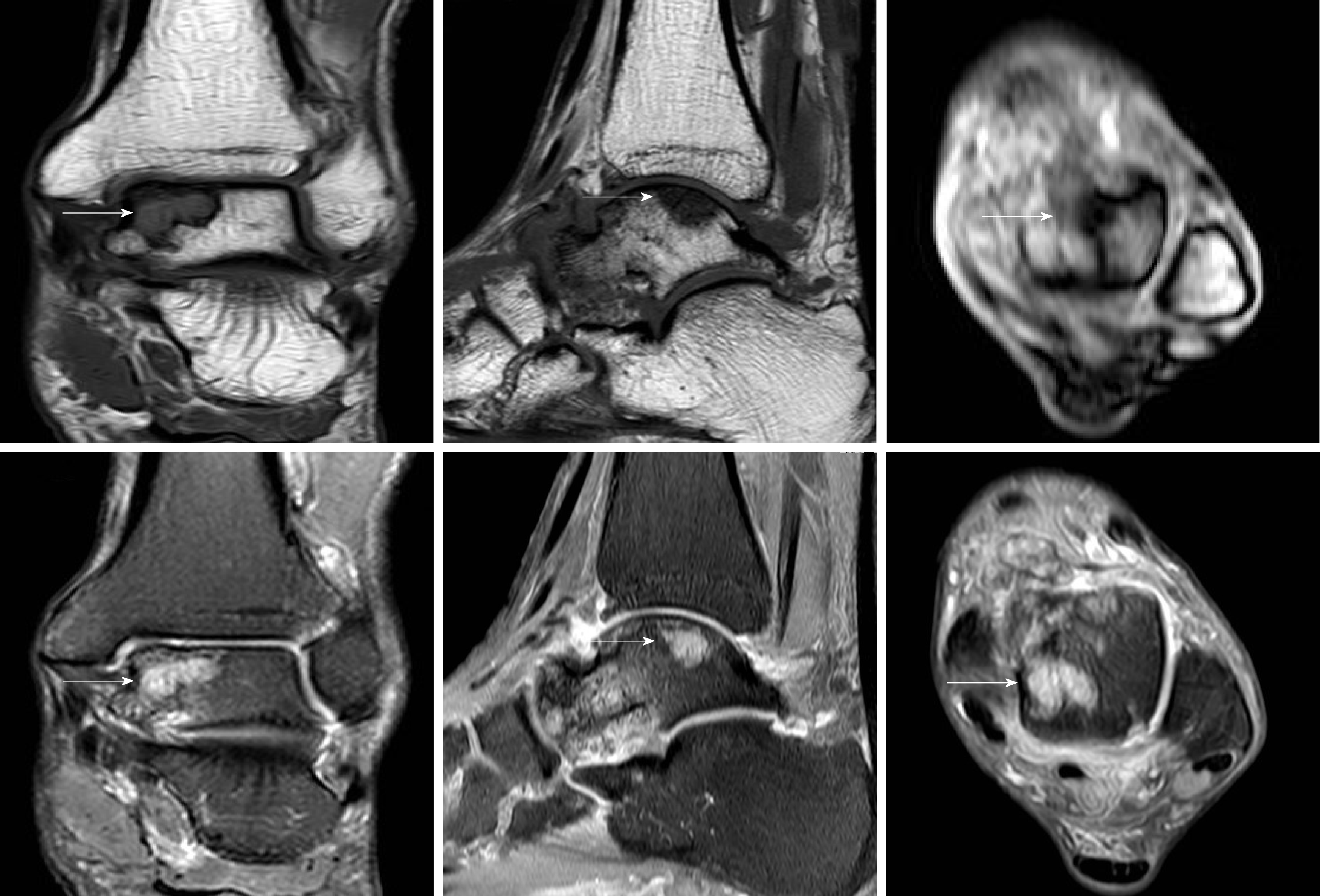
Figure 5 Magnetic resonance imaging showed osteolytic lesions in the talus hypointense on T1WI and hyperintense on T2WI (white arrow).
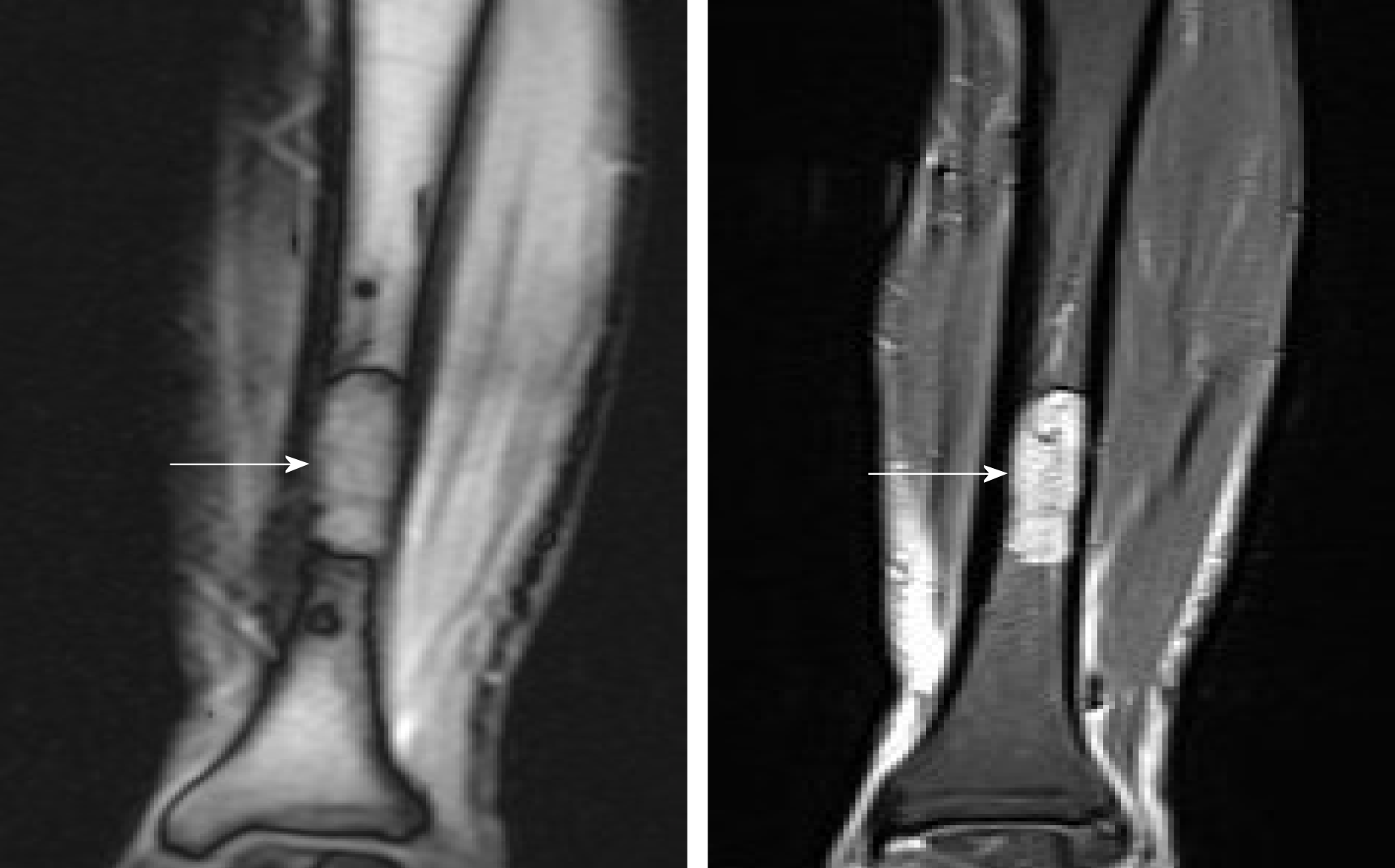
Figure 6 Magnetic resonance imaging suggested a marrow-replacing infiltrative lesion of the tibia hypointense on T1WI and hyperintense on T2WI.
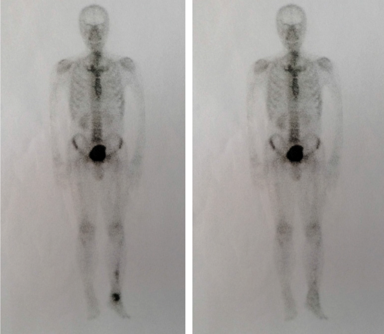
Figure 7 Comparison of preoperative and postoperative whole-body bone scans.
Figure 8 Computed tomography showed that lesions in the talus were completely removed and the bone was filled with cement casting.
Figure 9 Postoperative plain X-rays indicated complete lesion removal and no signs of recurrence.
- Citation: Xia Q, Tao C, Zhu KW, Zhong WY, Li PL, Jiang Y, Mao MZ. Erdheim-Chester disease with asymmetric talus involvement: A case report. World J Clin Cases 2020; 8(3): 614-623
- URL: https://www.wjgnet.com/2307-8960/full/v8/i3/614.htm
- DOI: https://dx.doi.org/10.12998/wjcc.v8.i3.614


