Copyright
©The Author(s) 2020.
World J Clin Cases. Feb 6, 2020; 8(3): 606-613
Published online Feb 6, 2020. doi: 10.12998/wjcc.v8.i3.606
Published online Feb 6, 2020. doi: 10.12998/wjcc.v8.i3.606
Figure 1 Pelvic contrast-enhanced computed tomography scan findings in our case of prostatic stromal sarcoma with rhabdoid features.
A: Axial computed tomography image showing the lesion as a heterogeneous mass, with areas of cystic change; B, C: Sagittal (B) and coronal (C) computed tomography images showing a large, heterogeneous mass arising from the prostate and invading the posterior wall of the bladder.
Figure 2 Microscopy findings in our case of prostatic stromal sarcoma with rhabdoid features.
A: Microphotograph showing the spindle cells arranged densely and loosely at intervals, with widespread myxoid background (H and E, × 10); B: The prostatic stroma showing the composition of spindle tumor cells with fascicular, whorled, and storiform arrangements (H and E, × 20); C: Areas of poor differentiation of embryonoid rhabdomyoblasts, with eccentric nuclei and unremarkable cross-striations, and showing abundant eosinophilic cytoplasm (H and E, × 100); D: Tumor cells showing the characteristics of nuclear division. Scattered distribution of prostatic glandular cells can be seen, and no malignant glandular epithelial cells were identified (H and E, × 60).
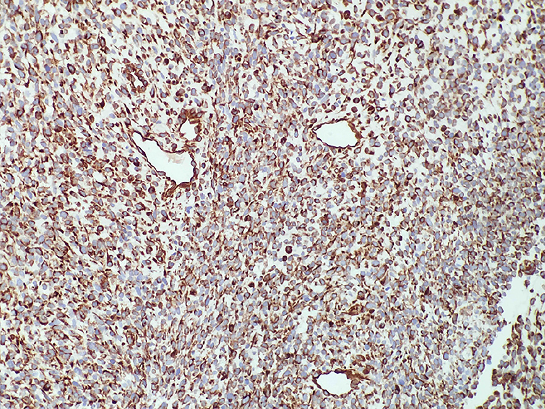
Figure 3 Angiomatoid type cells were present in the tumor in our case of prostatic stromal sarcoma with rhabdoid features.
Immunostaining of the tumor cells for vimentin was diffuse and strong (× 20).
Figure 4 Immunostaining results of our case of prostatic stromal sarcoma with rhabdoid features.
A, B: The rhabdomyoblasts stained positive for MyoD1 (A) and myogenin (B) (× 20); C: The tumor cells and rhabdomyoblasts stained positive for INI1 (× 20).
Figure 5 Additional immunostaining results in our case of prostatic stromal sarcoma with rhabdoid features.
A: The cytoplasm and cellular membrane of the spindle cells were immunoreactive for β-catenin, and the nuclei were unstained; B: Ki-67 staining; C: P53-positive spindle cells and glandular epithelial cells were found sporadically throughout the specimen; D: The glandular epithelial cells were positive for prostate-specific antigen.
- Citation: Li RG, Huang J. Clinicopathologic characteristics of prostatic stromal sarcoma with rhabdoid features: A case report. World J Clin Cases 2020; 8(3): 606-613
- URL: https://www.wjgnet.com/2307-8960/full/v8/i3/606.htm
- DOI: https://dx.doi.org/10.12998/wjcc.v8.i3.606


