Copyright
©The Author(s) 2020.
World J Clin Cases. Feb 6, 2020; 8(3): 600-605
Published online Feb 6, 2020. doi: 10.12998/wjcc.v8.i3.600
Published online Feb 6, 2020. doi: 10.12998/wjcc.v8.i3.600
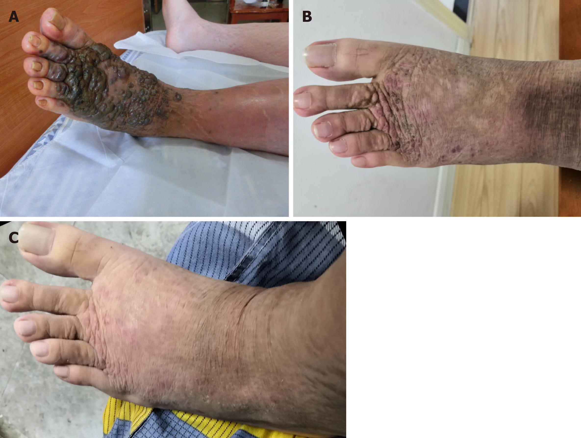
Figure 1 Cutaneous epithelioid angiomatous nodules appeared on admission and during the follow-up period.
A: 7 d; B: 10 mo; C: 16 mo.
Figure 2 Immunosuppressive treatment for the patient.
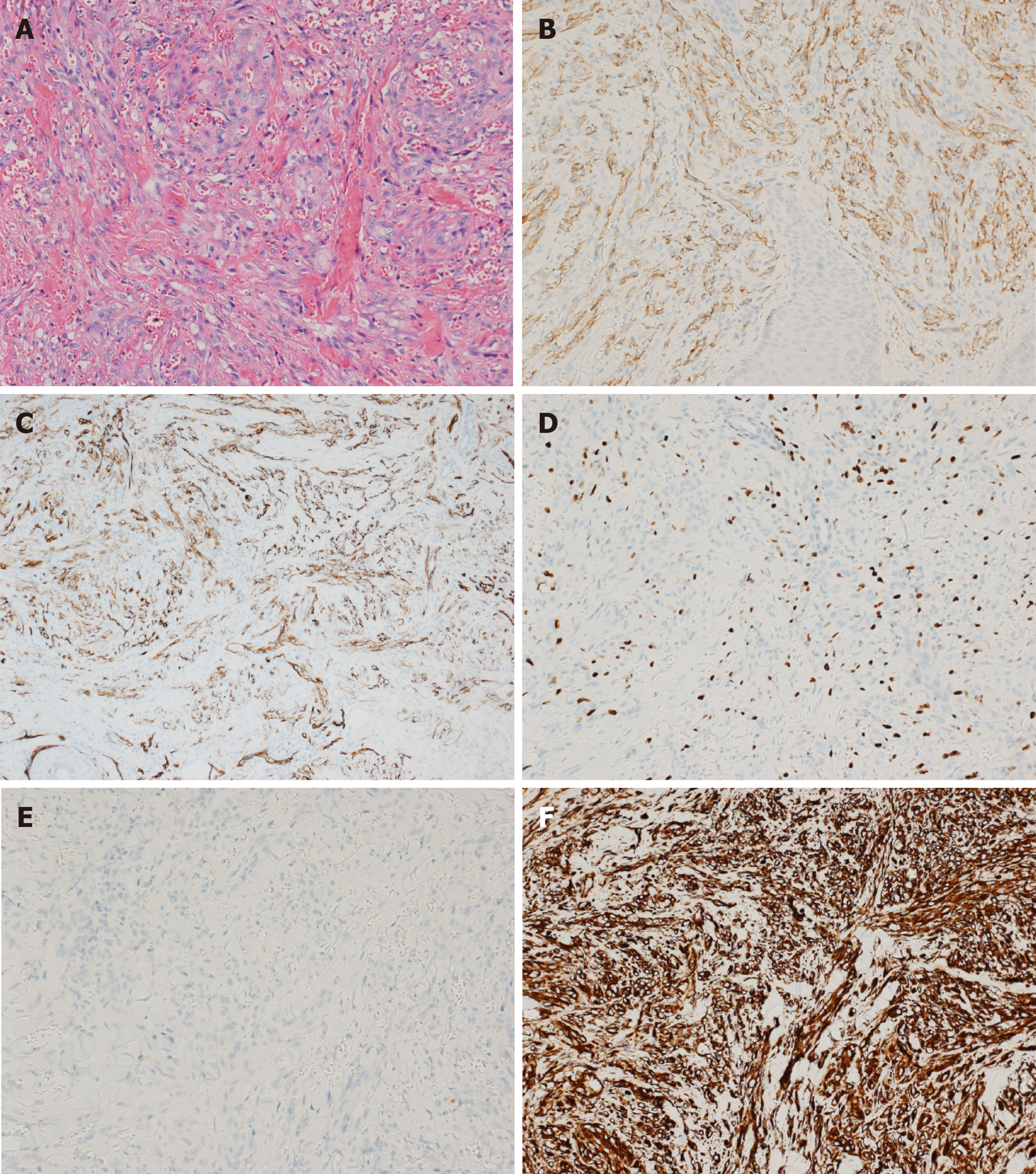
Figure 3 Histopathology.
A: Hematoxylin-eosin staining (× 200); B: Cluster of differentiation 31 positivity in endothelial cells (× 200); C: Cluster of differentiation 34 positivity in endothelial cells (× 200); D: Ki-67 (approximately 30%) positivity in endothelial cells (× 200); E: Cytokeratin negativity in endothelial cells (× 200); F: Vimentin negativity in endothelial cells (× 200).
- Citation: Cheng DJ, Zheng XY, Tang SF. Large cutaneous epithelioid angiomatous nodules in a patient with nephrotic syndrome: A case report. World J Clin Cases 2020; 8(3): 600-605
- URL: https://www.wjgnet.com/2307-8960/full/v8/i3/600.htm
- DOI: https://dx.doi.org/10.12998/wjcc.v8.i3.600


