Copyright
©The Author(s) 2020.
World J Clin Cases. Feb 6, 2020; 8(3): 552-559
Published online Feb 6, 2020. doi: 10.12998/wjcc.v8.i3.552
Published online Feb 6, 2020. doi: 10.12998/wjcc.v8.i3.552
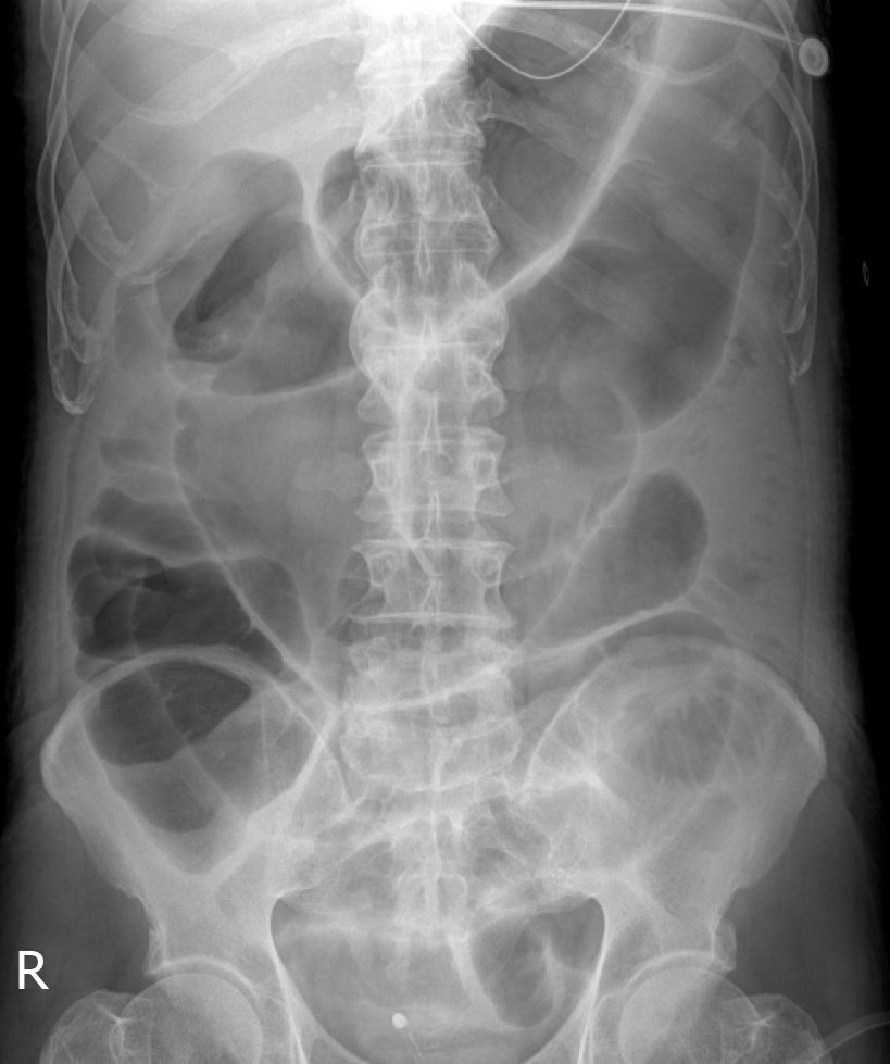
Figure 1 An X-ray image of the abdomen.
Abdominal film showing marked distensions of loops of the large and small intestines.
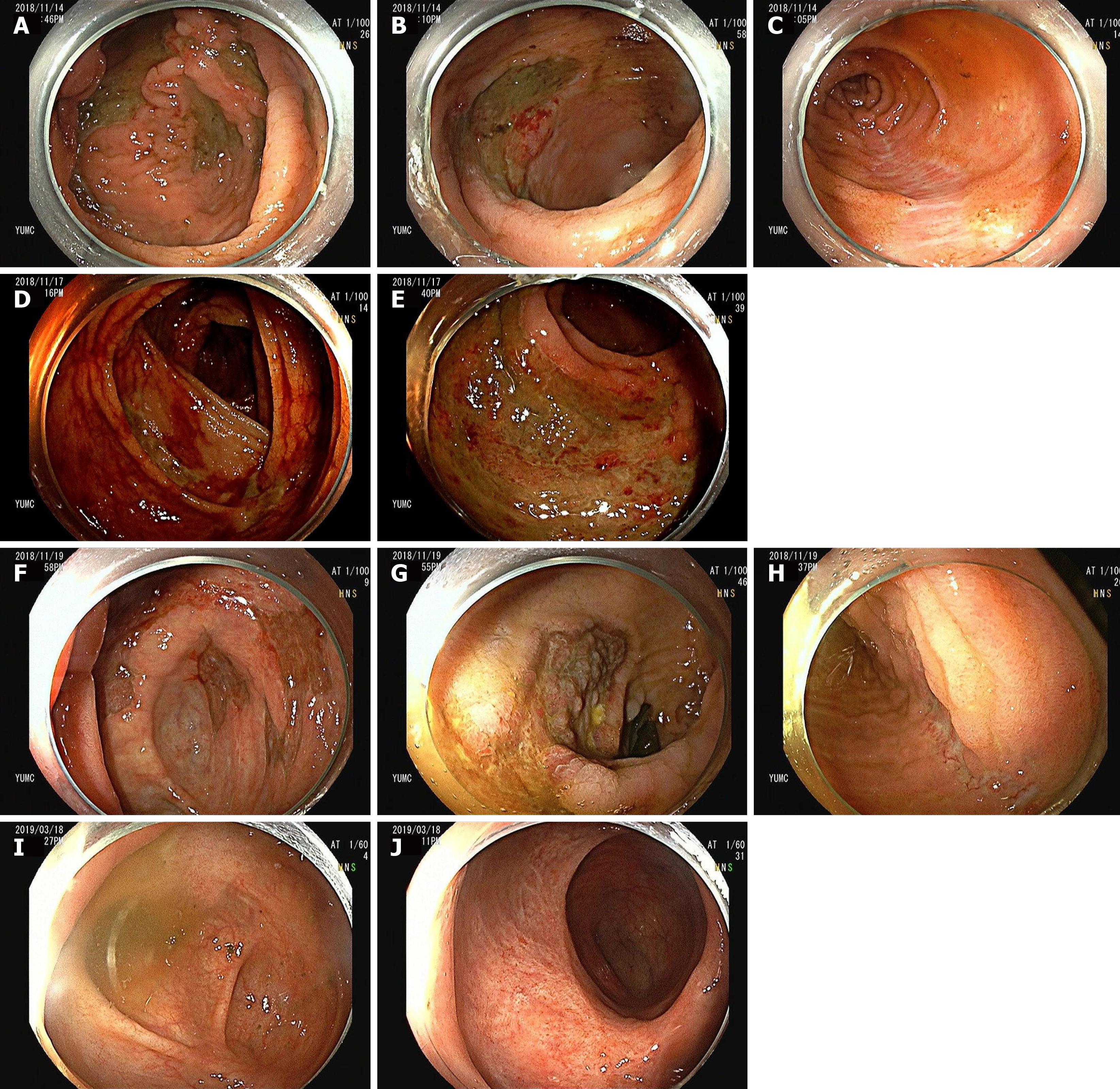
Figure 2 Colonoscopy images.
A-C: Colonoscopy images obtained on hospital day 8 (the next day of the first episode of massive hematochezia), showing huge, variably-sized, multiple, diffuse, deep ulcers at cecum (A) and sigmoid colon (B), and multiple healing stages ulcers at distal ileum (C); D, E: Follow-up colonoscopy images obtained on hospital day 10 showing oozing blood and multiple ulcers at ascending colon (D) and diffuse, huge ulcers at rectum (E); F-H: Follow-up colonoscopy images obtained on hospital day 14 (7 d after the first hematochezia event) showing huge and variably-sized, multiple, diffuse, healing stage ulcers with pinkish granulation tissue bases on cecum (F) and sigmoid colon (G), and multiple healing stage ulcers in distal ileum (H); I, J: Last follow-up colonoscopy image obtained after 2 mo of discharge showing several ulcer scars in cecum (I) and rectum (J) but no specific lesion.
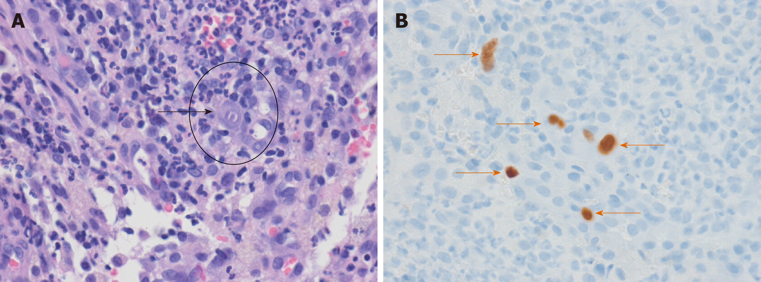
Figure 3 Pathology findings of hematoxylin-eosin and immunohistochemical stained biopsy sections.
A: The black arrow shows cytomegalovirus inclusion bodies (HE staining, × 400); B: Orange arrows show cytomegalovirus-positive cells (immunohistochemical staining, × 400).
- Citation: Cho JH, Choi JH. Cytomegalovirus ileo-pancolitis presenting as toxic megacolon in an immunocompetent patient: A case report. World J Clin Cases 2020; 8(3): 552-559
- URL: https://www.wjgnet.com/2307-8960/full/v8/i3/552.htm
- DOI: https://dx.doi.org/10.12998/wjcc.v8.i3.552


