Copyright
©The Author(s) 2020.
World J Clin Cases. Oct 6, 2020; 8(19): 4633-4643
Published online Oct 6, 2020. doi: 10.12998/wjcc.v8.i19.4633
Published online Oct 6, 2020. doi: 10.12998/wjcc.v8.i19.4633
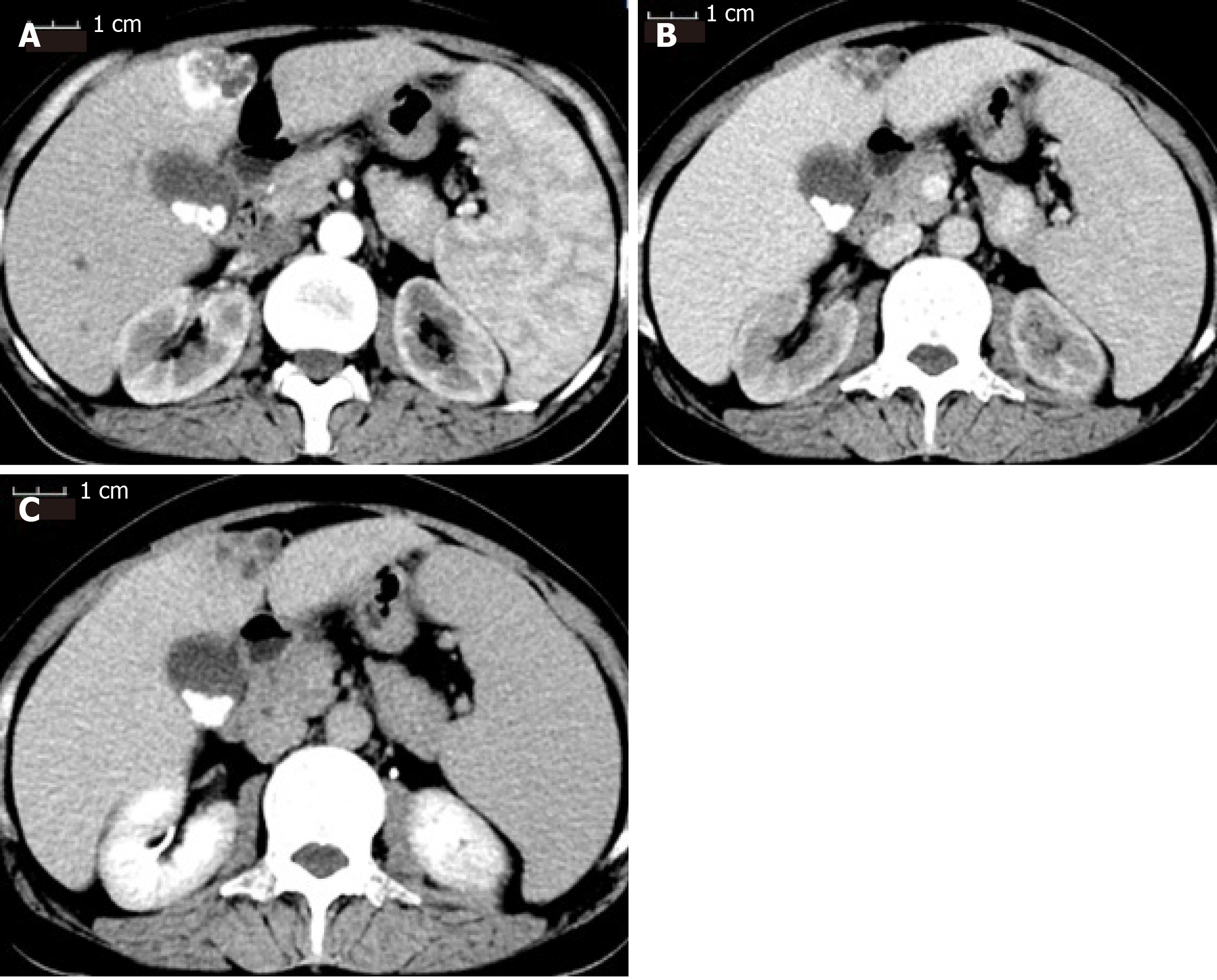
Figure 1 Contrast-enhanced computed tomography scan of the patient before receiving transarterial chemoembolization.
A: The mass was enhanced immediately during the arterial phase; B: The enhancement faded away quickly into equal density in the portal venous phase; C: The mass showed hypodensity in the delayed phase. The three phases of the computed tomography scan also showed multiple gallstones and splenomegaly.
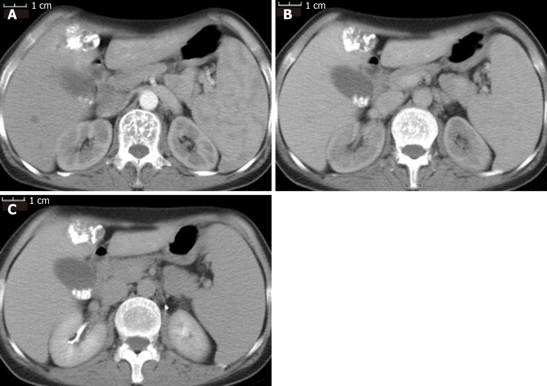
Figure 2 Contrast-enhanced computed tomography scan of the patient after first transarterial chemoembolization.
A: Iodized oil deposition in the tumor was observed in the arterial phase; B: Portal venous phase; C: Delayed phase.
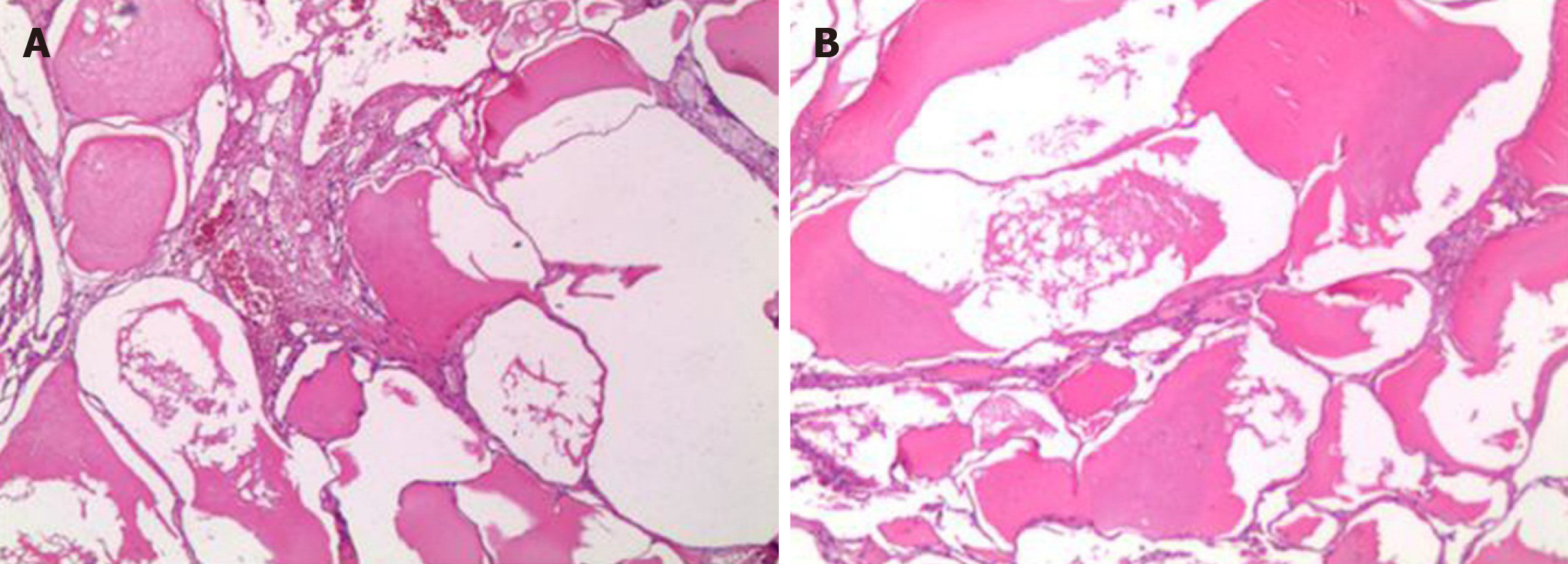
Figure 3 The specimen was examined under microscopy (40 ×).
A: The mass was made of dilated-cystic lymphatic lumens lined by endothelium; B: Filled with acidophilic lymph.
- Citation: Long X, Zhang L, Cheng Q, Chen Q, Chen XP. Solitary hepatic lymphangioma mimicking liver malignancy: A case report and literature review. World J Clin Cases 2020; 8(19): 4633-4643
- URL: https://www.wjgnet.com/2307-8960/full/v8/i19/4633.htm
- DOI: https://dx.doi.org/10.12998/wjcc.v8.i19.4633


