Copyright
©The Author(s) 2020.
World J Clin Cases. Oct 6, 2020; 8(19): 4535-4543
Published online Oct 6, 2020. doi: 10.12998/wjcc.v8.i19.4535
Published online Oct 6, 2020. doi: 10.12998/wjcc.v8.i19.4535
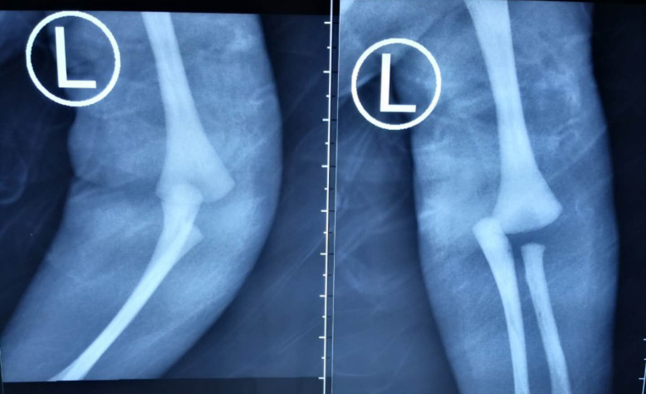
Figure 1 Anteroposterior and lateral radiographs of the elbow joint from the primary hospital, indicating dislocation of the elbow joint.
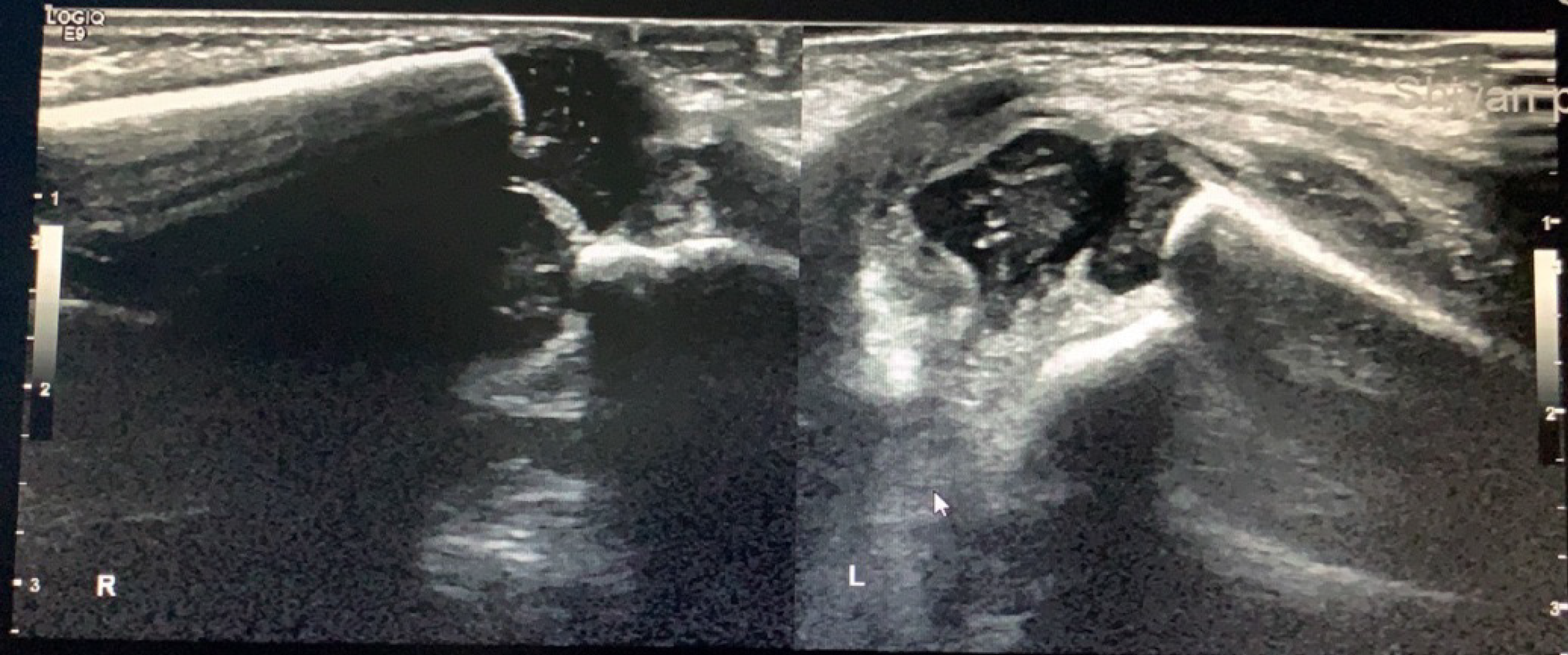
Figure 2 Ultrasonograms of the elbow joint from the primary hospital, indicating physeal fracture of the left distal humerus.
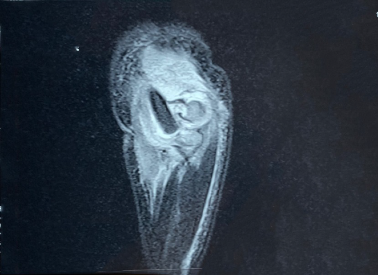
Figure 3 Magnetic resonance imaging of the elbow joint from the primary hospital, indicating physeal fracture of the left distal humerus without callus formation.
Figure 4 On admission, physical examination revealed considerable swelling of the elbow, tenderness, and upper limb dysfunction.
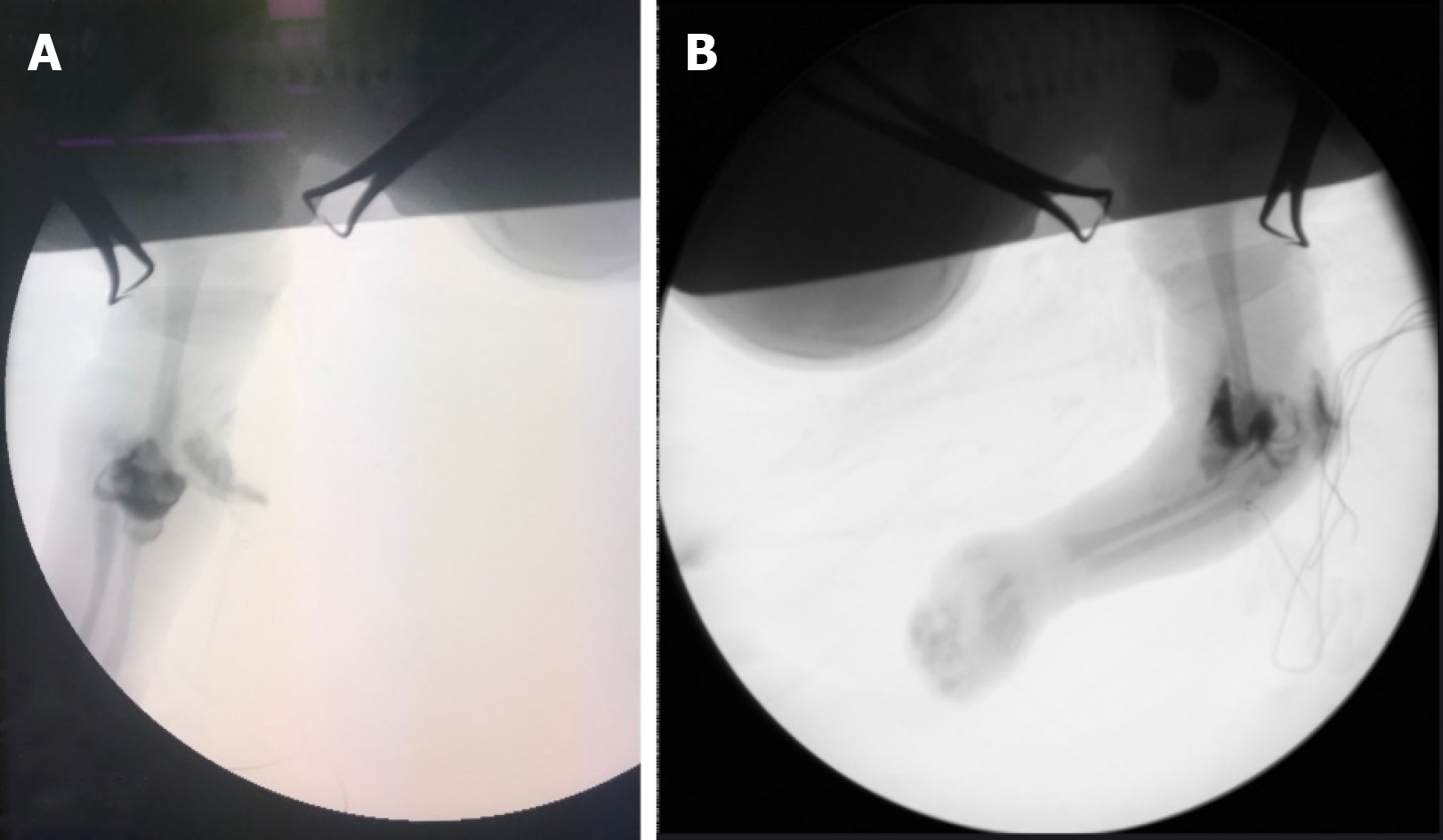
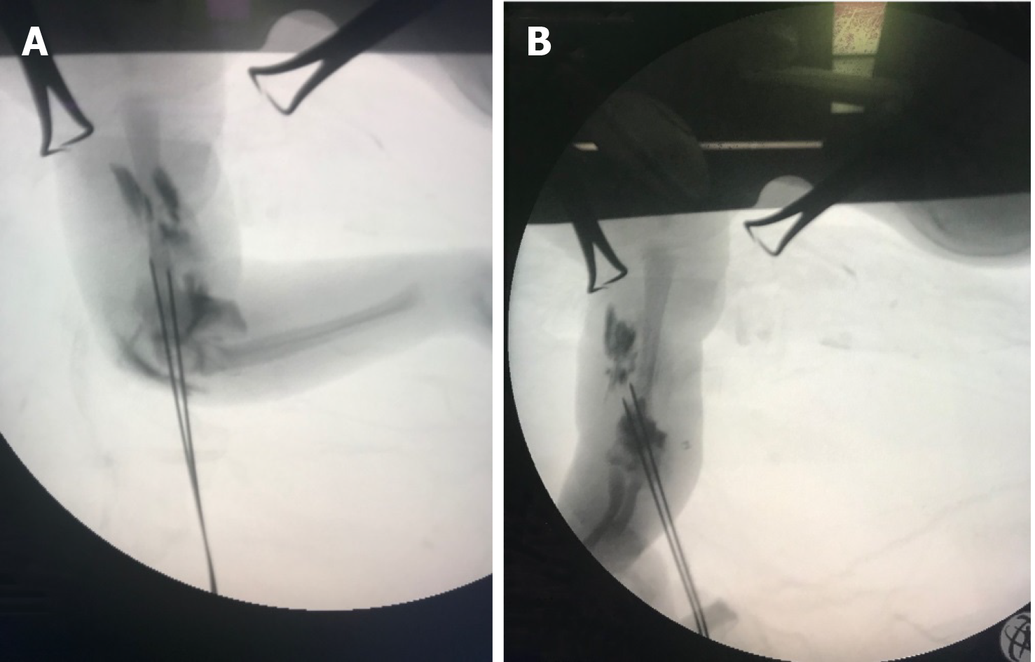
Figure 5 Arthrography of the elbow during the operation revealed physeal fracture of the distal humerus, and posteromedial displacement of the distal epiphysis (A and B).
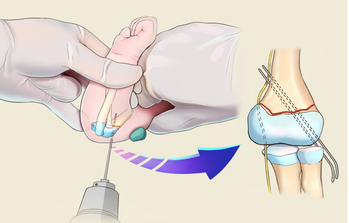
Figure 6 Sketch of manual reduction.
Figure 7 Internal fixation was performed using Kirschner wire (A and B).
Subsequent arthrography revealed satisfactory reduction of the fracture.
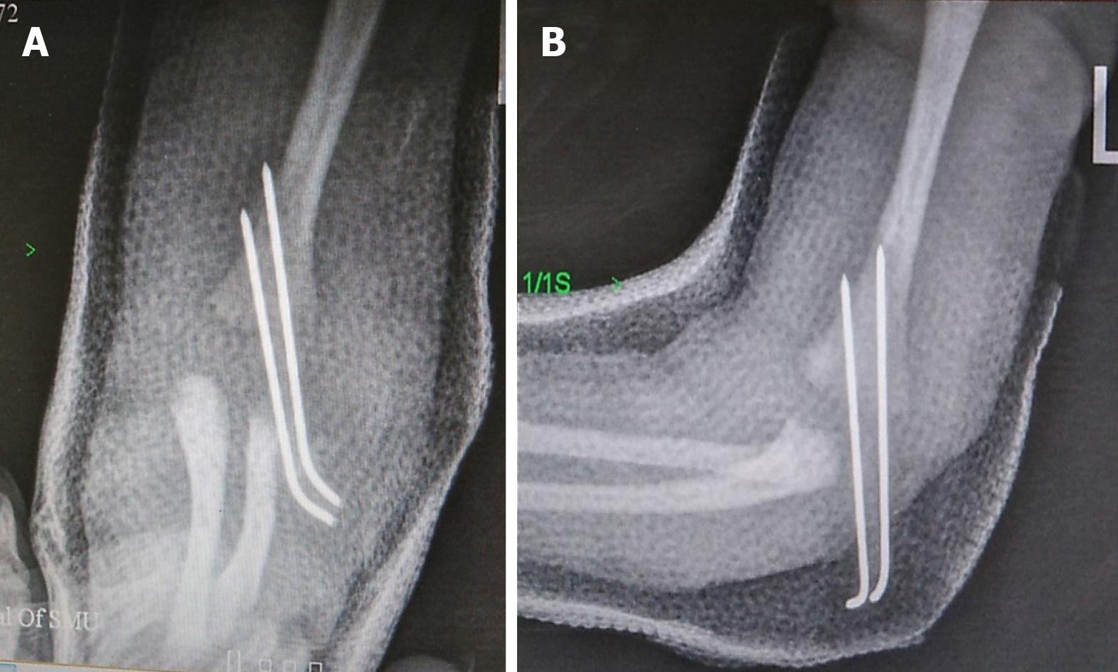
Figure 8 Postoperative anteroposterior and lateral radiographs of the elbow joint indicated the alignment of the humeral-ulnar joint with satisfactory reduction and fixation of the physeal fracture of the distal humerus (A and B).
- Citation: Tan W, Wang FH, Yao JH, Wu WP, Li YB, Ji YL, Qian YP. Percutaneous fixation of neonatal humeral physeal fracture: A case report and review of the literature. World J Clin Cases 2020; 8(19): 4535-4543
- URL: https://www.wjgnet.com/2307-8960/full/v8/i19/4535.htm
- DOI: https://dx.doi.org/10.12998/wjcc.v8.i19.4535


