Copyright
©The Author(s) 2020.
World J Clin Cases. Oct 6, 2020; 8(19): 4505-4511
Published online Oct 6, 2020. doi: 10.12998/wjcc.v8.i19.4505
Published online Oct 6, 2020. doi: 10.12998/wjcc.v8.i19.4505
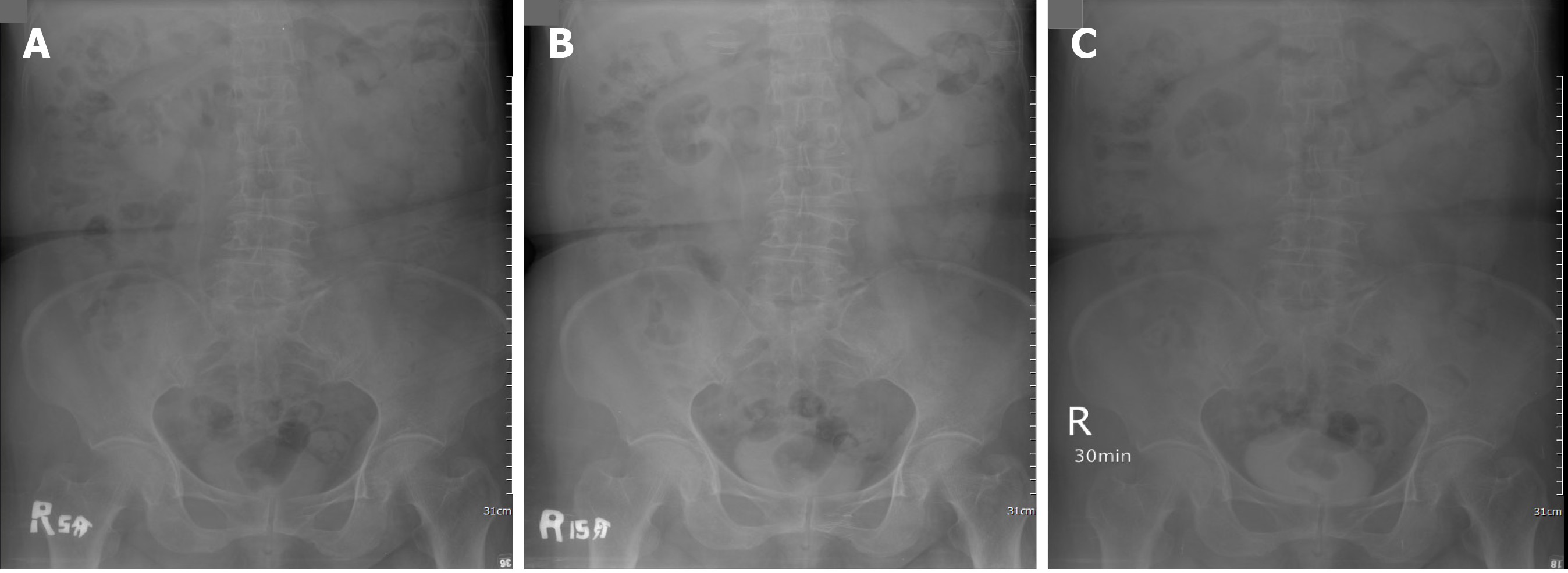
Figure 1 Intravenous pyelography.
No enhancement of urinary tract at 5 min (A), 15 min (B) and 30 min (C) after administration of the contrast.
Figure 2 Computed tomography urogram.
One 4.2 cm irregular mass was seen occupying the left renal pelvis on computed tomography urogram (A) (B); One enlarged lymph node was observed at the left renal pedicle (C).
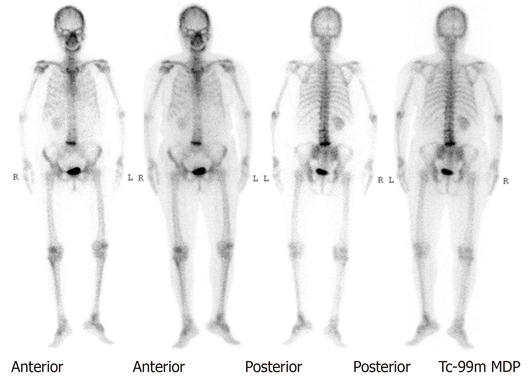
Figure 3 No observed active lesions were demonstrated on whole-body bone scan.
Figure 4 Hematoxylin and eosin stain.
A: Generally, with hematoxylin and eosin stain the tumor was seen with invasion to muscular layer; B and C: Under augmentation (400 ×), the tumor was examined with epithelial cells and lymphoid cells.
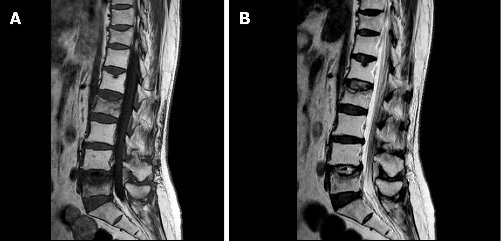
Figure 5 Magnetic resonance imaging.
T1-weight (A) and T2-weight (B) image revealed destructive L2 and L5 spine. Related involvement of nearby spinal cord was also noted.
- Citation: Yang CH, Weng WC, Lin YS, Huang LH, Lu CH, Hsu CY, Ou YC, Tung MC. Eight-year follow-up of locally advanced lymphoepithelioma-like carcinoma at upper urinary tract: A case report. World J Clin Cases 2020; 8(19): 4505-4511
- URL: https://www.wjgnet.com/2307-8960/full/v8/i19/4505.htm
- DOI: https://dx.doi.org/10.12998/wjcc.v8.i19.4505


