Copyright
©The Author(s) 2020.
World J Clin Cases. Oct 6, 2020; 8(19): 4499-4504
Published online Oct 6, 2020. doi: 10.12998/wjcc.v8.i19.4499
Published online Oct 6, 2020. doi: 10.12998/wjcc.v8.i19.4499
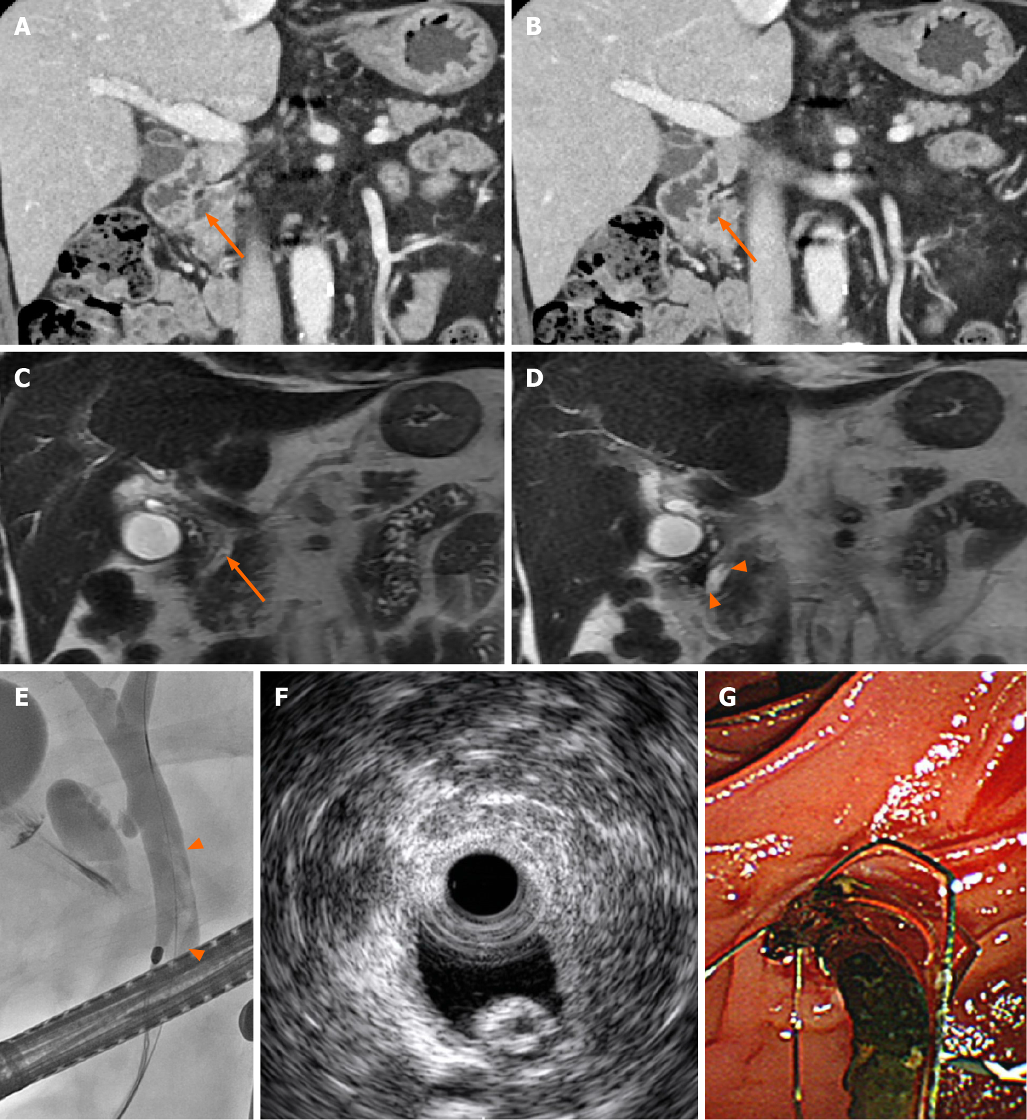
Figure 1 A 72-year-old male with a distal common bile duct stone mimicking Ascaris.
A and B: Coronal reformatted images of contrast-enhanced computed tomography showed a hyperattenuated, elongated lesion inside the distal common bile duct (CBD) lumen (arrows); C and D: A T2-weighted coronal image revealed a linear filling defect in the distal CBD lumen with mild ductal dilatation, findings that mimic an Ascaris worm; E and F: An elongated tubular filling defect was seen on endoscopic retrograde cholangiopancreatography, and a hyperechoic lesion with a central hypoechoic area was seen on endoscopic ultrasound; G: A dark-brown-colored CBD stone was discovered through endoscopic removal.
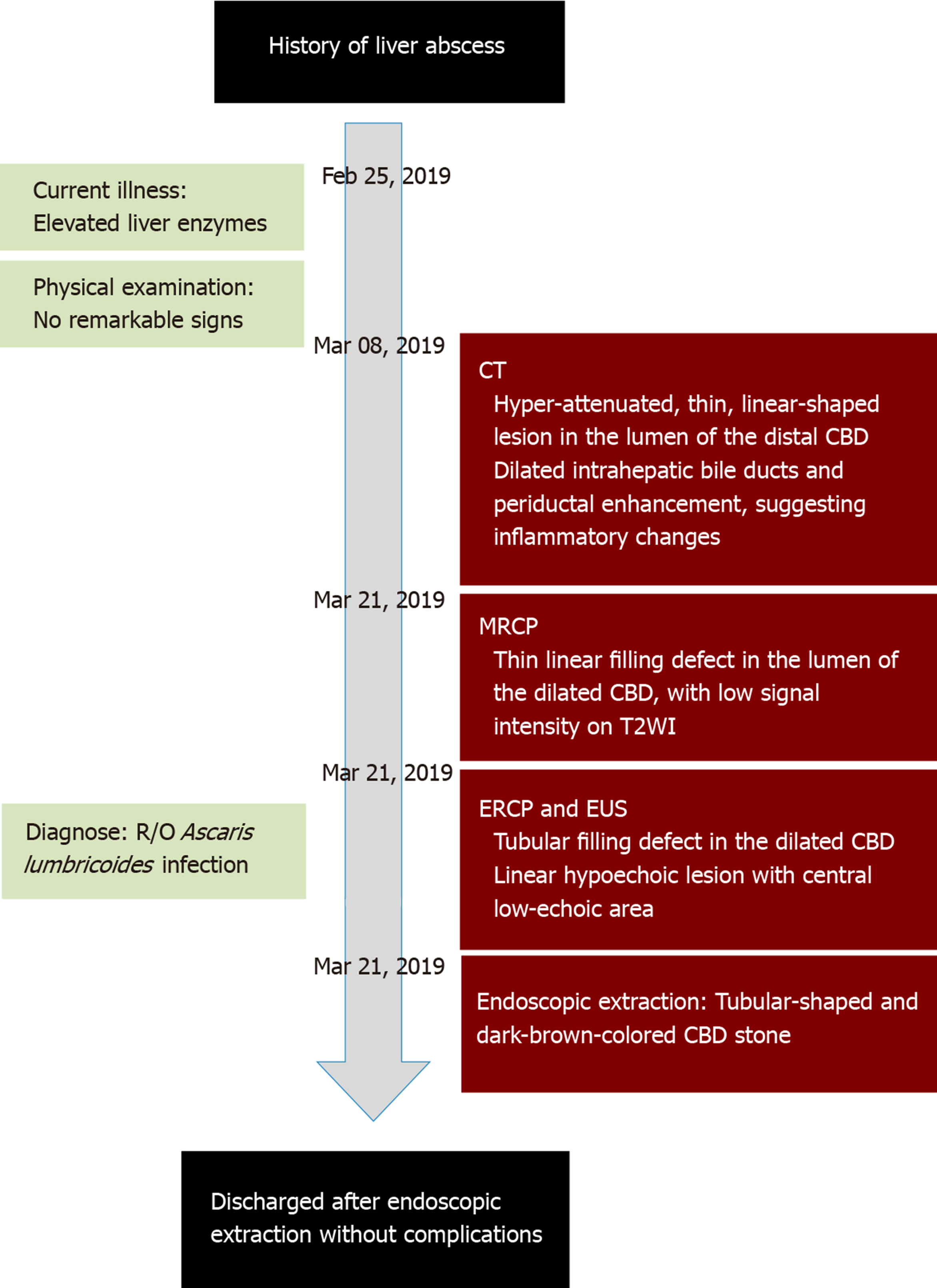
Figure 2 Timeline of the patient’s illness.
CT: Computed tomography; CBD: Common bile duct; MRCP: Magnetic resonance cholangiopancreatography; ERCP: Endoscopic retrograde cholangiopancreatography; EUS: Endoscopic ultrasound.
- Citation: Choi SY, Jo HE, Lee YN, Lee JE, Lee MH, Lim S, Yi BH. Ascaris-mimicking common bile duct stone: A case report. World J Clin Cases 2020; 8(19): 4499-4504
- URL: https://www.wjgnet.com/2307-8960/full/v8/i19/4499.htm
- DOI: https://dx.doi.org/10.12998/wjcc.v8.i19.4499


