Copyright
©The Author(s) 2020.
World J Clin Cases. Oct 6, 2020; 8(19): 4481-4487
Published online Oct 6, 2020. doi: 10.12998/wjcc.v8.i19.4481
Published online Oct 6, 2020. doi: 10.12998/wjcc.v8.i19.4481
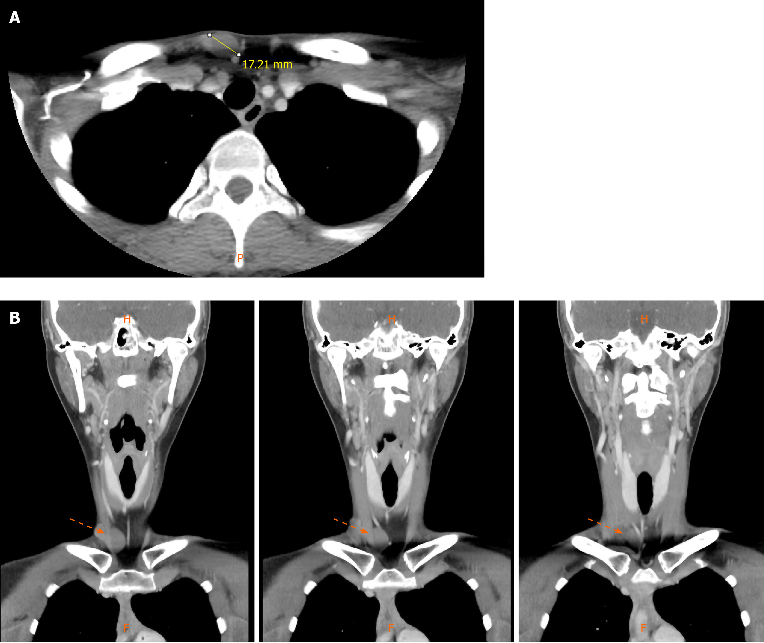
Figure 1 Enhanced computed tomography of the neck.
A: Non-enhancing round mass on the subcutaneous area in the anterior neck area in axial view; B: Serial images of coronal section.
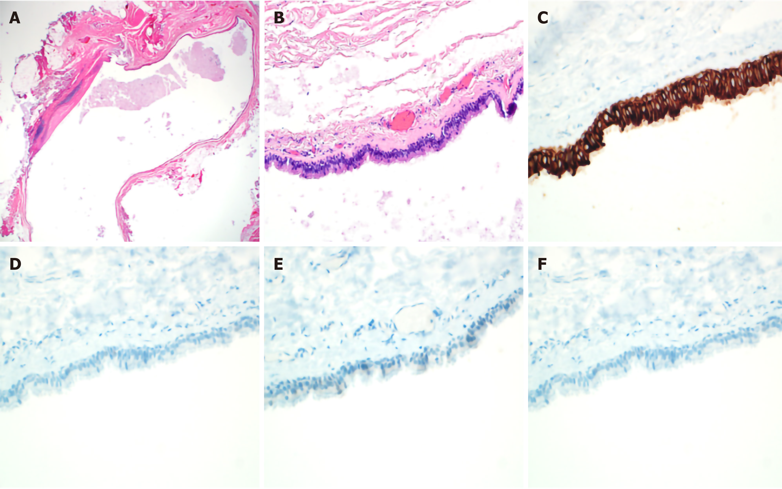
Figure 2 Pathologic findings reveal a well-defined ovoid unilocular cystic lesion lined by ciliated pseudostratified cuboidal to columnar epithelium.
A and B: Focal intraluminal papillary projections in the subcutaneous tissue (Hematoxylin and eosin stain, × 400); C: The lining epithelium reveals immunoreactive cytokeratin (cytokeratin, × 400); D-F: It reveals negative findings for immunostainings (D: Estrogen receptor, × 400; E: Desmin, × 400; F: S-100, × 400).
- Citation: Kim YH, Lee J. Cutaneous ciliated cyst on the anterior neck in young women: A case report. World J Clin Cases 2020; 8(19): 4481-4487
- URL: https://www.wjgnet.com/2307-8960/full/v8/i19/4481.htm
- DOI: https://dx.doi.org/10.12998/wjcc.v8.i19.4481


