Copyright
©The Author(s) 2020.
World J Clin Cases. Oct 6, 2020; 8(19): 4443-4449
Published online Oct 6, 2020. doi: 10.12998/wjcc.v8.i19.4443
Published online Oct 6, 2020. doi: 10.12998/wjcc.v8.i19.4443
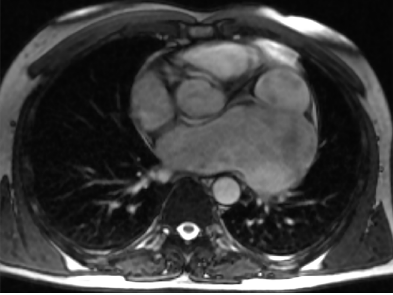
Figure 1 Magnetic resonance imaging.
A white arrow shows a sharply dilated cavity of the left atrial appendage.
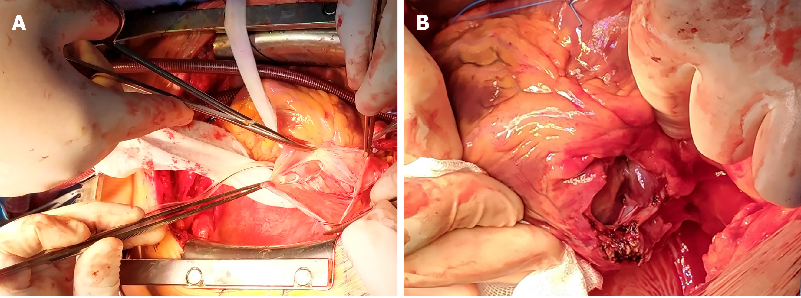
Figure 2 Aneurysmectomy.
A: The aneurysm of the left atrial appendage is opened; B: The aneurysm of the left atrial appendage is excised and sutured from the inside.
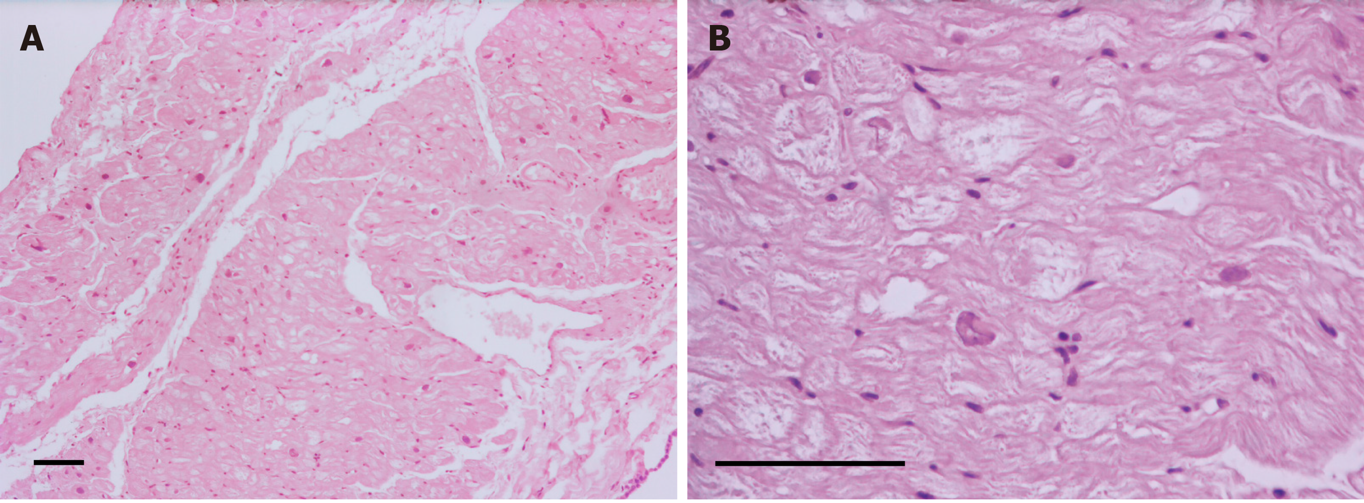
Figure 3 Hematoxylin and eosin staining of the resected left atrial appendage aneurysm.
A: The wall of the aneurysm is thin, with slight fibrosis of the endo- and epicardium (scale bar 100 µm, 100 × magnification); B: Hypertrophied cardiomyocytes with dystrophic changes and vacuolated cytoplasm (scale bar 100 µm, 400 × magnification).
- Citation: Belov DV, Moskalev VI, Garbuzenko DV, Arefyev NO. Left atrial appendage aneurysm: A case report. World J Clin Cases 2020; 8(19): 4443-4449
- URL: https://www.wjgnet.com/2307-8960/full/v8/i19/4443.htm
- DOI: https://dx.doi.org/10.12998/wjcc.v8.i19.4443


