Copyright
©The Author(s) 2020.
World J Clin Cases. Oct 6, 2020; 8(19): 4286-4302
Published online Oct 6, 2020. doi: 10.12998/wjcc.v8.i19.4286
Published online Oct 6, 2020. doi: 10.12998/wjcc.v8.i19.4286
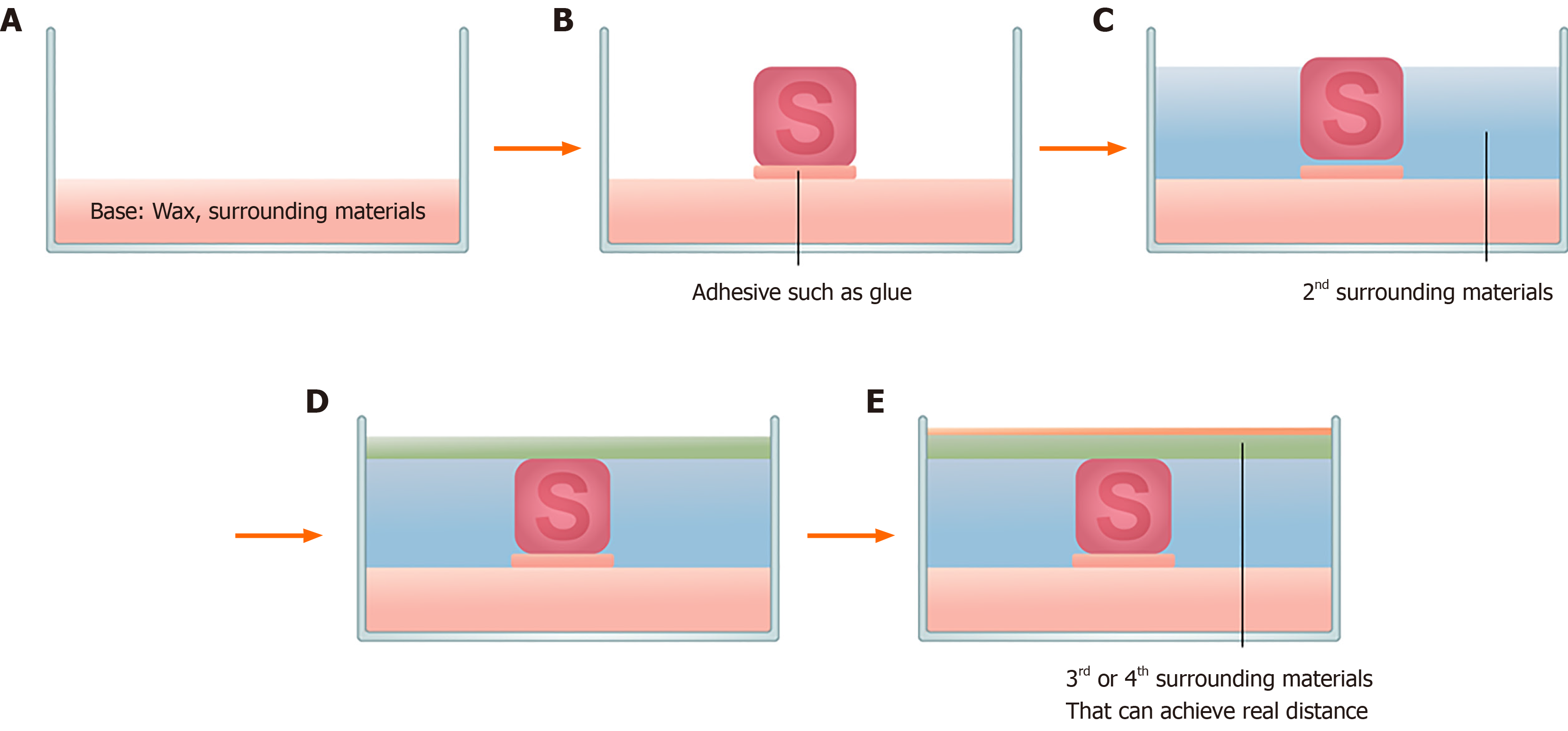
Figure 1 General method to produce the simulator.
The surrounding material and specific simulator are used in a container box. A: The surrounding material or wax is used to form a base in a container box prior to the specific simulator insertion; B: When the base material solidifies, the specific simulator is glued with an adhesive; C: The process of inserting the surrounding material; D and E: The surrounding material can be inserted at once or may be split into two depending on the purpose of the project. At this time, air bubble is removed using a spoon.
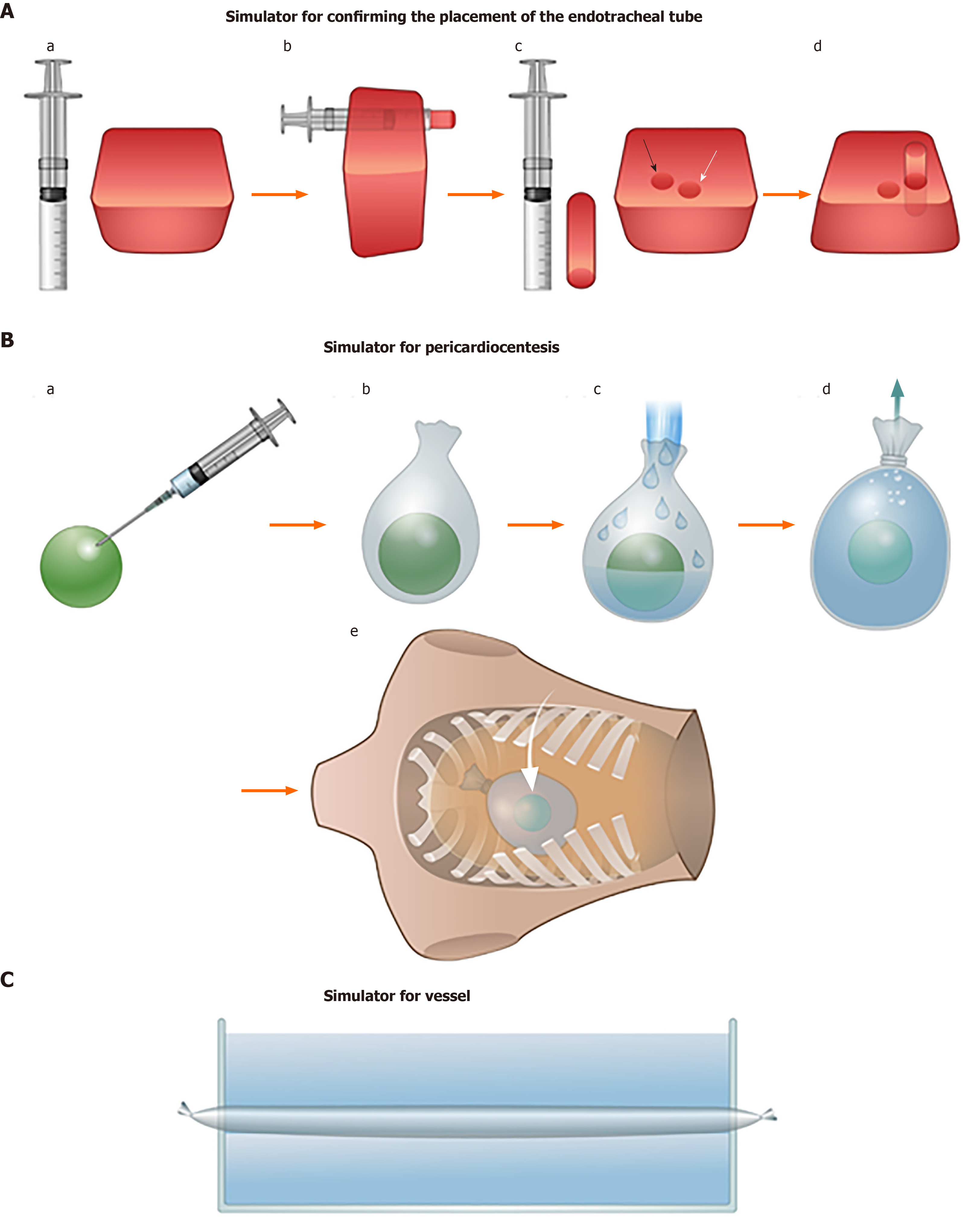
Figure 2 S specific simulator: The process of making some specific simulator that is complicated to make.
A: Simulator for confirming the placement of the endotracheal tube A (a) beef gelatin powder (90 mL) and 60 mL of orange-colored psyllium fiber (Metamucil sugar free; P and G, Cincinnati, OH, United States) mixed with 500 mL of boiling water in a 1 L container. A 10 mL syringe with the tip cutoff; A (b) The two holes are separated by a gap between the trachea and oesophagus; they are 5 mm from the wall of the margins of the surrounding materials to achieve a real sonographic appearance. Two holes were made using a 10 mL syringe with the tip cutoff once the surrounding material was completely ready; A (c) White arrow shows the trachea. Black arrow shows the oesophagus; A (d) The simulation with the block in the hole that represented the oesophagus did not show the double tracheal sign and showed tracheal intubation. B: Pericardiocentesis simulator B (a) Prick a hole in a ping-pong ball and fill with water. It represents a heart; B (b) The ball then is inserted into a balloon or a 250 cc saline bag, which represents the pericardium; B (c) The balloon is filled with water to show the pericardial fluid; B (d) A spoon or a syringe is used to remove the bubbles; B (e) the simulator is used in an order as in Figure 1, with an artificial rib cage or a dummy. The posterior portion of the artificial rib cage and posterior portion of a dummy are removed so that it can be used as a container. And C: Vessel simulator such as latex or balloon are inserted in process Figure 1.
- Citation: Shin KC, Ha YR, Lee SJ, Ahn JH. Review of simulation model for education of point-of-care ultrasound using easy-to-make tools. World J Clin Cases 2020; 8(19): 4286-4302
- URL: https://www.wjgnet.com/2307-8960/full/v8/i19/4286.htm
- DOI: https://dx.doi.org/10.12998/wjcc.v8.i19.4286


