Copyright
©The Author(s) 2020.
World J Clin Cases. Sep 26, 2020; 8(18): 4266-4271
Published online Sep 26, 2020. doi: 10.12998/wjcc.v8.i18.4266
Published online Sep 26, 2020. doi: 10.12998/wjcc.v8.i18.4266
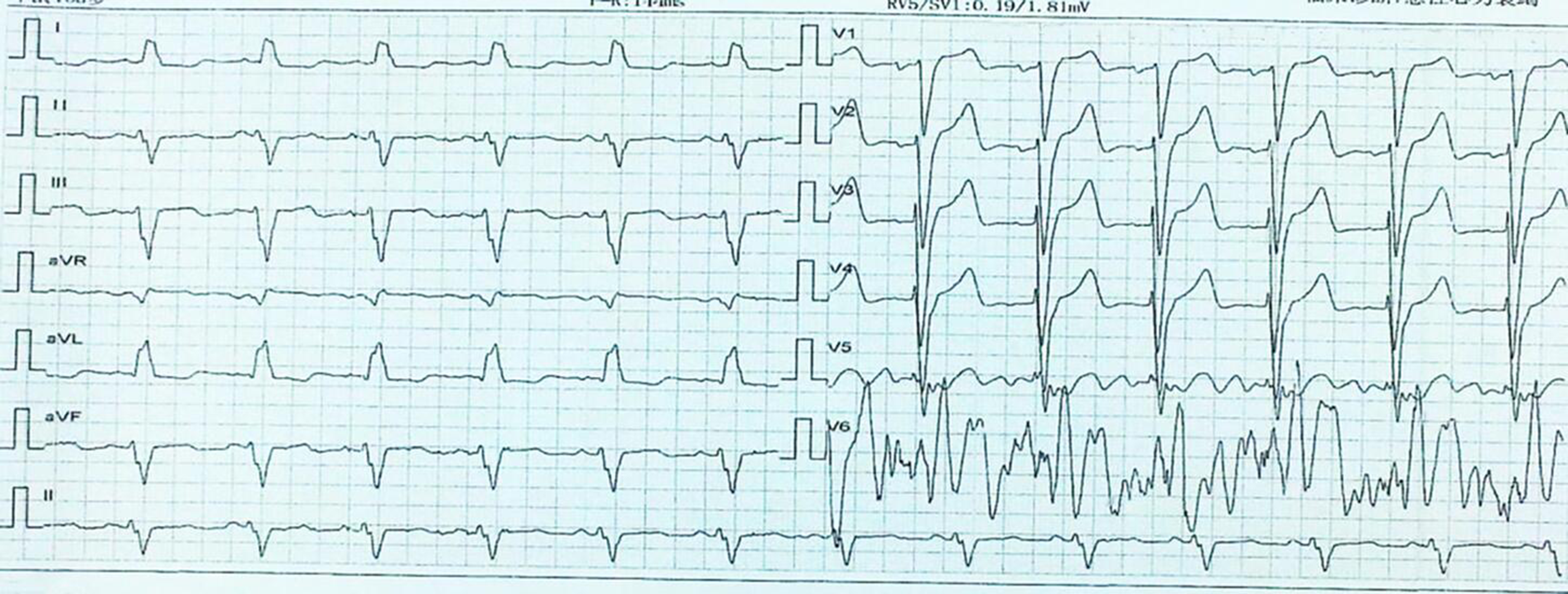
Figure 1 Preoperative basic electrocardiogram.
The electrocardiogram results showed sinus rhythm, complete left bundle branch block, and QRS duration of 160 ms.
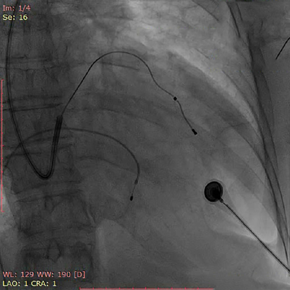
Figure 2 Electrode image.
The left ventricular wire was successfully inserted into the left ventricular vein of the coronary sinus.
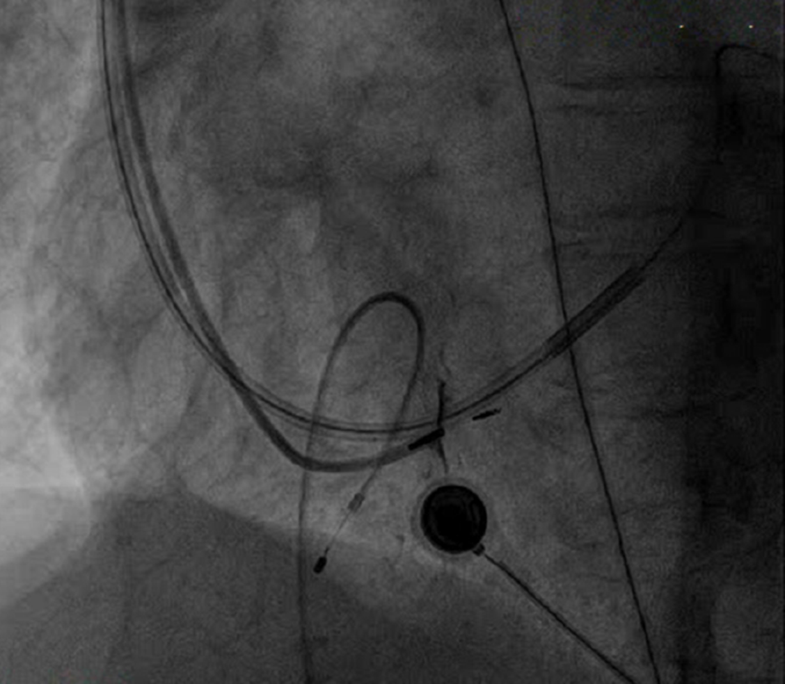
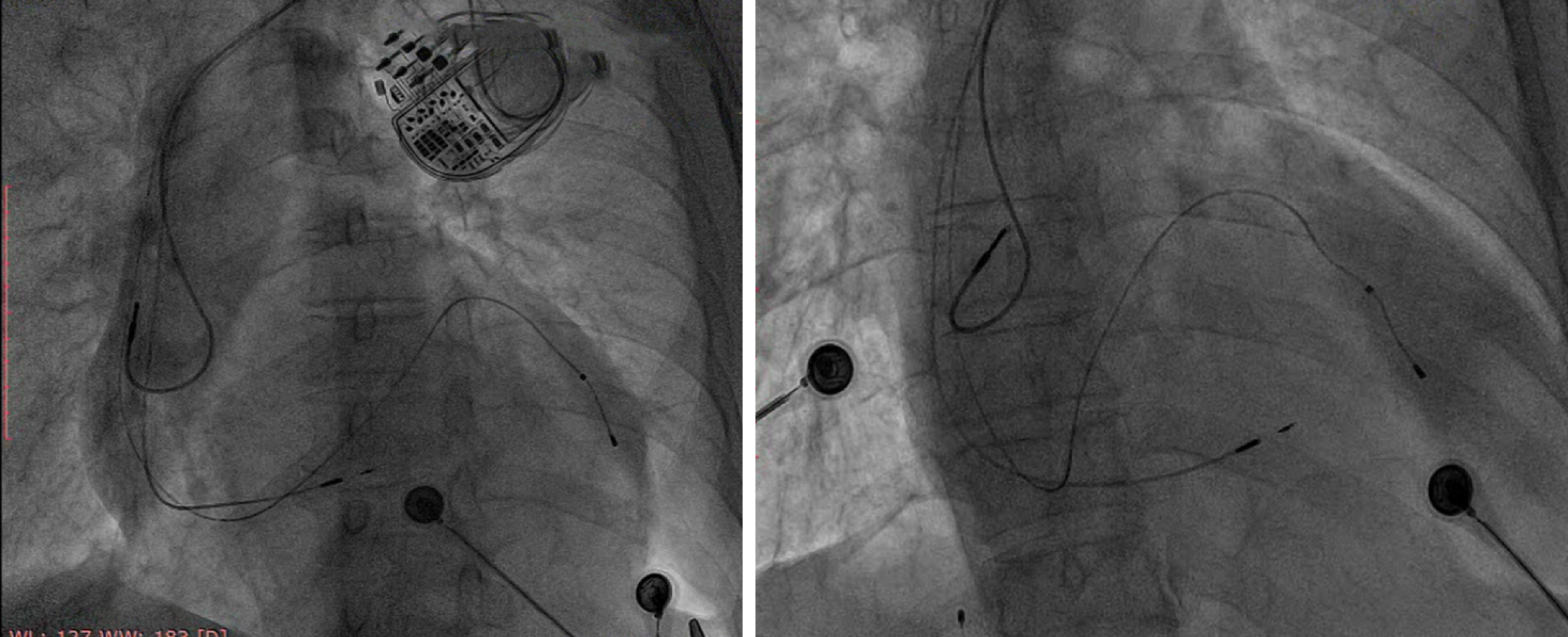
Figure 3 Electrode image.
The wire was placed in the region of the left bundle branch, and the depth of the electrode could be seen.
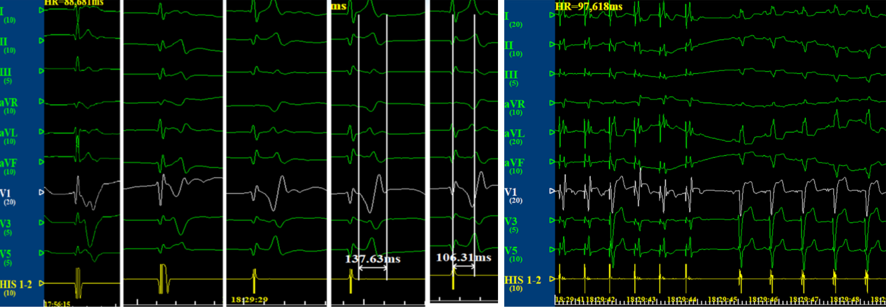
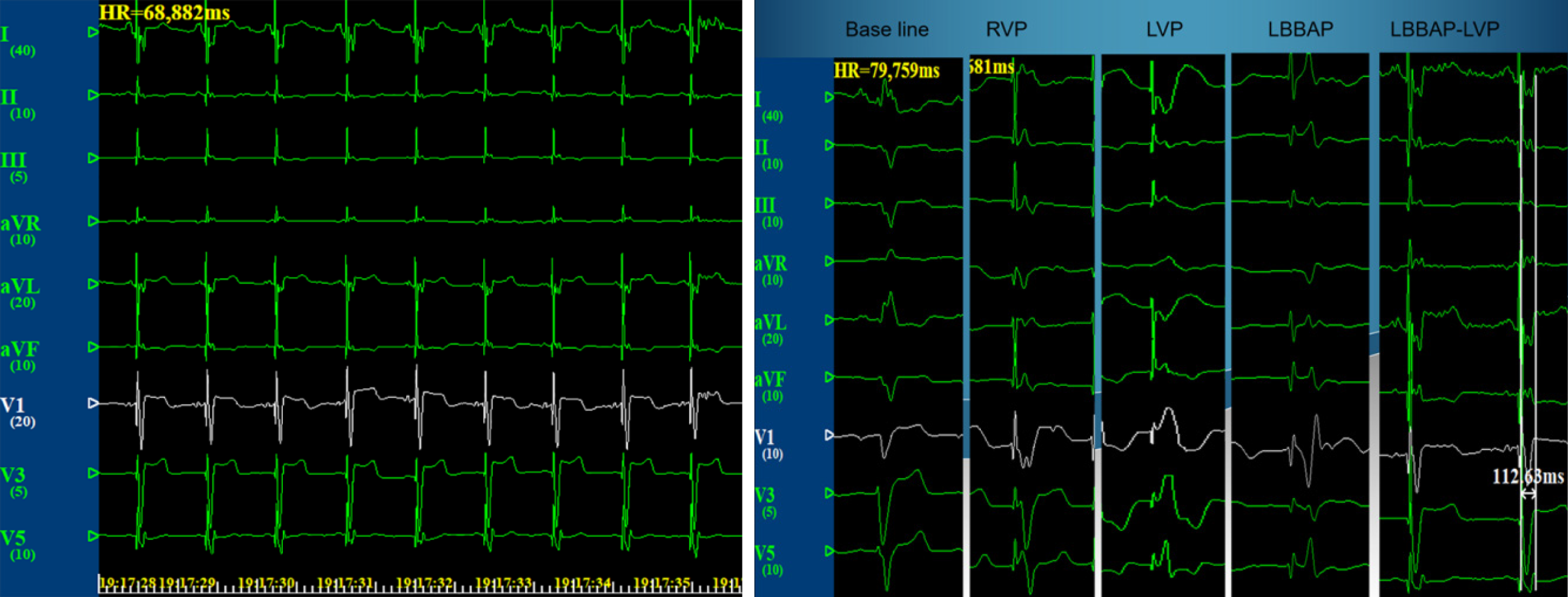
Figure 4 Electrocardiogram.
Unipolar pacing of the right ventricular septum and gradual rotation of the interior septum to reach the left bundle branch region (lead V1 R wave gradually increased, and the final QRS wave width was 137 ms). Comparison of the correction of complete left bundle branch block with unipolar pacing using ECG.
Figure 5 Electrocardiogram.
A: Graph when the atrioventricular delay was adjusted to 130 ms, and the two-chamber pacemaker was fused with the right bundle branch and down transmission (QRS 110 ms); B: Left bundle branch pacing with the optimization of cardiac resynchronization.
Figure 6 Final electrode images (left anterior oblique position: 30 degrees; posterior and anterior sides).
- Citation: Zhang DH, Lang MJ, Tang G, Chen XX, Li HF. Left bundle branch pacing with optimization of cardiac resynchronization treatment: A case report. World J Clin Cases 2020; 8(18): 4266-4271
- URL: https://www.wjgnet.com/2307-8960/full/v8/i18/4266.htm
- DOI: https://dx.doi.org/10.12998/wjcc.v8.i18.4266


