Copyright
©The Author(s) 2020.
World J Clin Cases. Sep 6, 2020; 8(17): 3875-3880
Published online Sep 6, 2020. doi: 10.12998/wjcc.v8.i17.3875
Published online Sep 6, 2020. doi: 10.12998/wjcc.v8.i17.3875
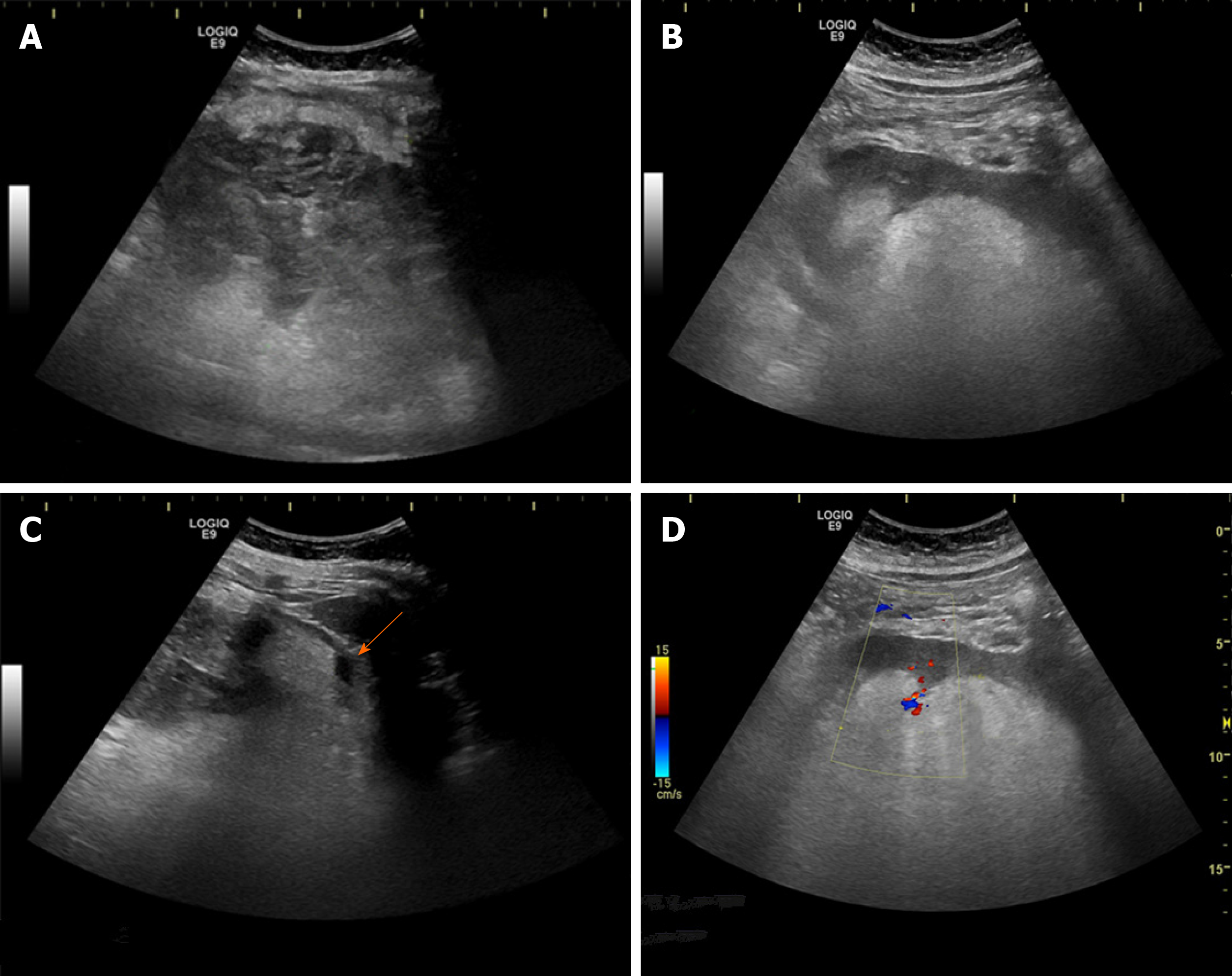
Figure 1 Ultrasound images of a giant nonhomogenous lump in the left kidney area.
A: The part of the lump located in the left kidney area was hypoechoic with a strong stripe echo; B: The lump caused the left kidney to move to the middle abdomen. The inner part of the lump near the left kidney was hyperechoic; C: A stripe-shaped echoless zone was seen around the lump (arrow); D: The color Doppler flow image showed some spot-like blood flow signals around the lump.
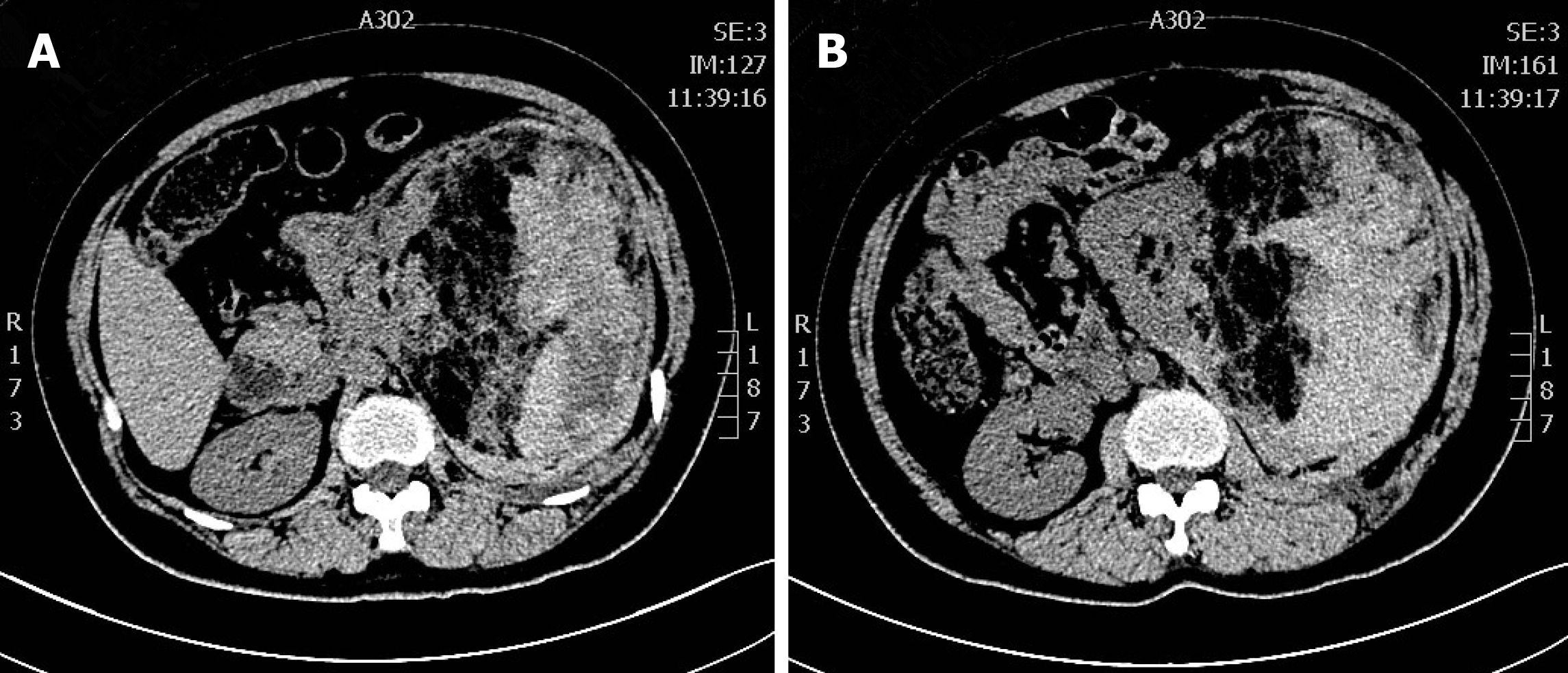
Figure 2 Computed tomography exhibited a large, mixed-density mass in the left kidney.
The density of the area adjacent to the kidney was low (A), and the area far from the kidney showed high density (B).
- Citation: Zhang T, Xue S, Wang ZM, Duan XM, Wang DX. Diagnostic value of ultrasound in the spontaneous rupture of renal angiomyolipoma during pregnancy: A case report. World J Clin Cases 2020; 8(17): 3875-3880
- URL: https://www.wjgnet.com/2307-8960/full/v8/i17/3875.htm
- DOI: https://dx.doi.org/10.12998/wjcc.v8.i17.3875


