Copyright
©The Author(s) 2020.
World J Clin Cases. Sep 6, 2020; 8(17): 3847-3852
Published online Sep 6, 2020. doi: 10.12998/wjcc.v8.i17.3847
Published online Sep 6, 2020. doi: 10.12998/wjcc.v8.i17.3847
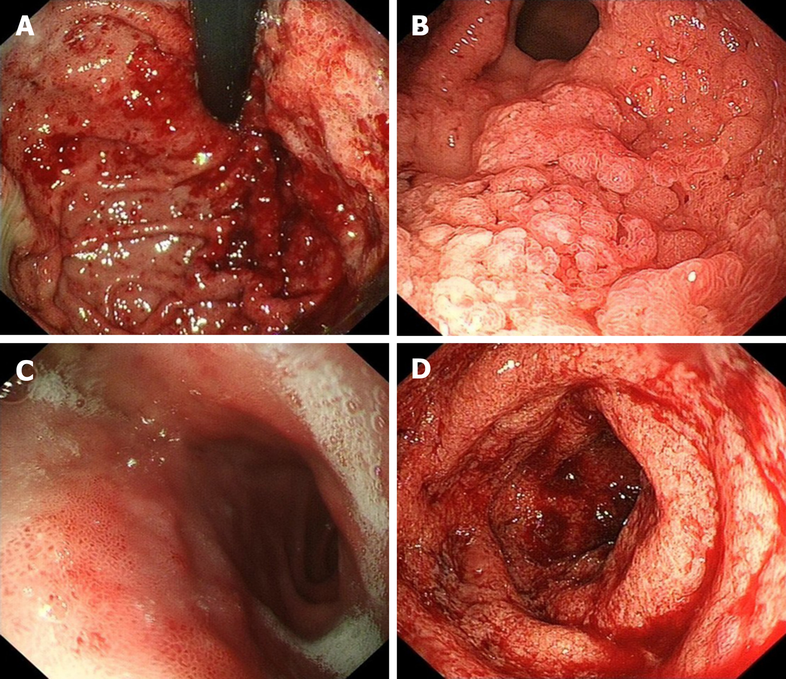
Figure 1 Mucosal manifestation under gastroscopy and enteroscopy.
A: Gastroscopy showed diffuse edema and local mucosal erosion, ulcers, and bleeding in the fundus of the stomach; B: Granular and friable changes were observed in the gastric antrum without causing lumen stenosis; C: The mucosa contained edema and erosions in front of the duodenal bulb; D: Continuous superficial ulcers and spontaneous bleeding were observed in the rectum by colonoscopy.
Figure 2 Biopsy specimens from the gastroduodenum and colorectum [hematoxylin and eosin (HE) staining, original magnification ×100].
A: Diffuse inflammation accompanied by an abscess in the lesser curvature of the stomach; B: Diffuse inflammation in the duodenum; C: Diffuse colitis with a crypt abscess.
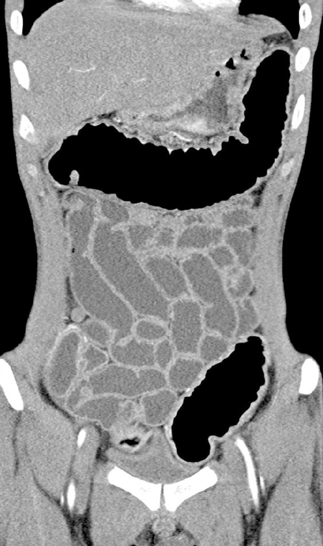
Figure 3 Abdominal computed tomography.
Diffuse thickening in stomach and colorectum wall was seen, while the small intestine was not involved.
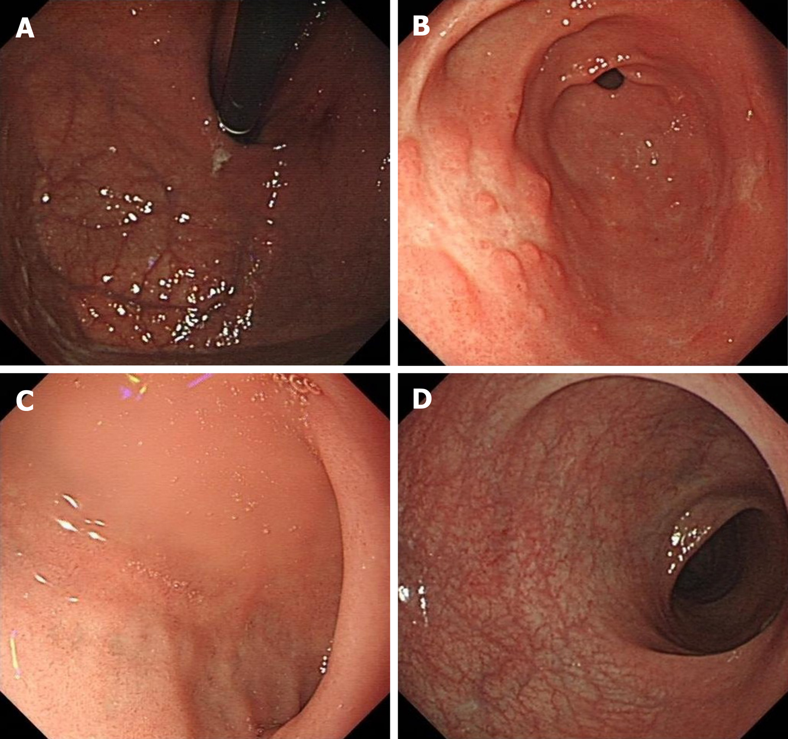
Figure 4 Follow-up endoscopy.
Endoscopy showed no significant abnormalities in the fundus of the stomach (A), duodenum (C ) or rectum (D). Polypoid hyperplasia was observed in the gastric antrum (B).
- Citation: Yang Y, Li CQ, Chen WJ, Ma ZH, Liu G. Gastroduodenitis associated with ulcerative colitis: A case report. World J Clin Cases 2020; 8(17): 3847-3852
- URL: https://www.wjgnet.com/2307-8960/full/v8/i17/3847.htm
- DOI: https://dx.doi.org/10.12998/wjcc.v8.i17.3847


