Copyright
©The Author(s) 2020.
World J Clin Cases. Sep 6, 2020; 8(17): 3808-3813
Published online Sep 6, 2020. doi: 10.12998/wjcc.v8.i17.3808
Published online Sep 6, 2020. doi: 10.12998/wjcc.v8.i17.3808
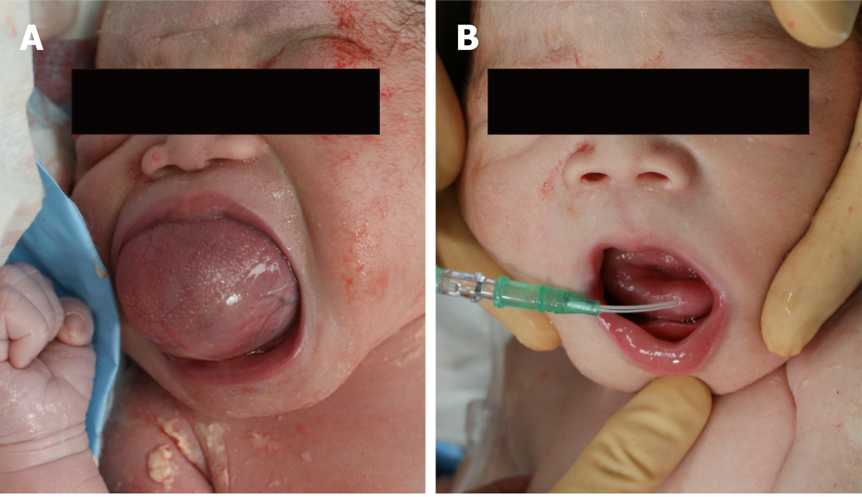
Figure 1 Neonatal appearance at birth.
A: The tongue cyst occupies the oral cavity before needle aspiration; B: Tongue cyst aspiration with an 18-gauge indwelling needle.
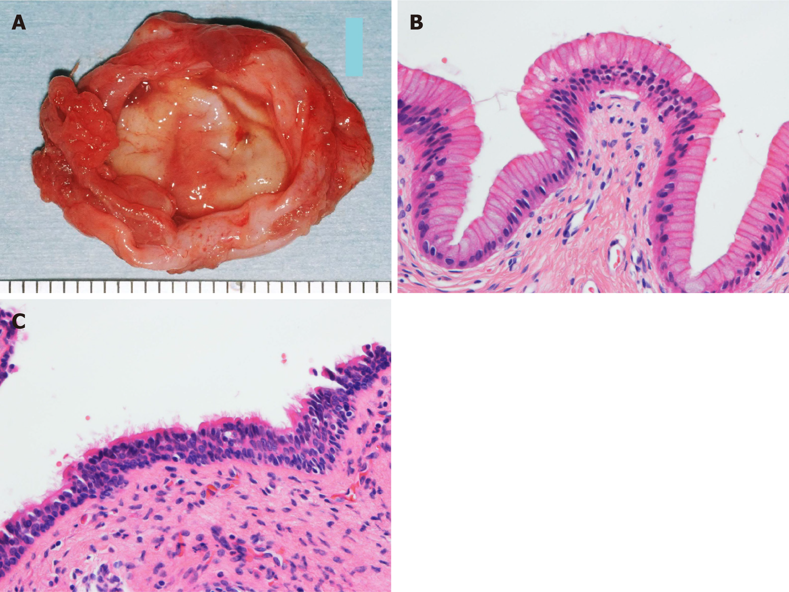
Figure 2 Pathological findings of the cyst.
A: Gross anatomy of the excised cyst. The cyst wall is elastic and soft, and the lumen is smooth; B: Hematoxylin-eosin stained specimen showing columnar gastrointestinal epithelium lining the cyst wall (× 400 original magnification); C: Ciliated pseudostratified epithelium is also seen in another part (× 400 original magnification).
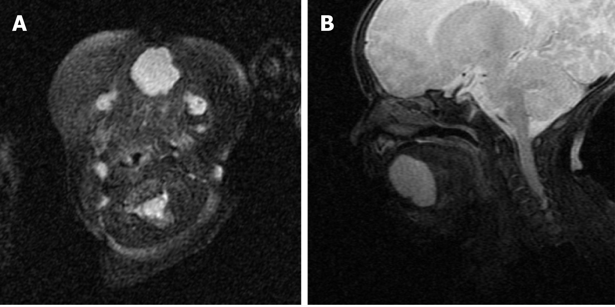
Figure 3 T2-weighted magnetic resonance imaging shows that the cyst is located at the ventral part of the anterior tongue.
A: Horizontal view; B: Sagittal view.
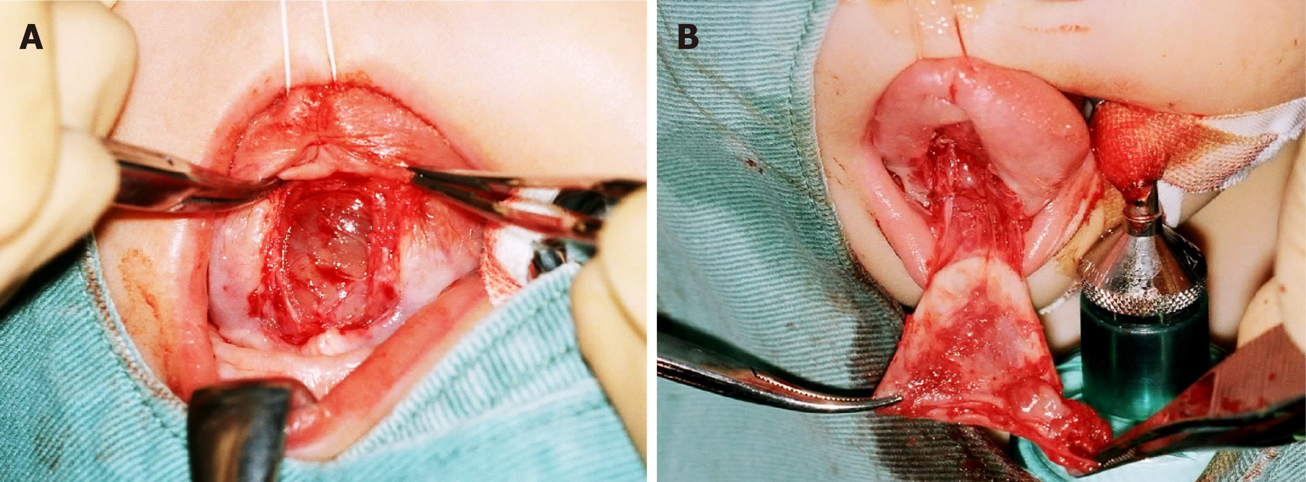
Figure 4 Intraoperative findings.
A: A smooth cyst wall is observed after dissecting the tongue mucosa; B: Loose connective tissue is attached to the cyst.
- Citation: Lee AD, Harada K, Tanaka S, Yokota Y, Mima T, Enomoto A, Kogo M. Large lingual heterotopic gastrointestinal cyst in a newborn: A case report. World J Clin Cases 2020; 8(17): 3808-3813
- URL: https://www.wjgnet.com/2307-8960/full/v8/i17/3808.htm
- DOI: https://dx.doi.org/10.12998/wjcc.v8.i17.3808


