Copyright
©The Author(s) 2020.
World J Clin Cases. Sep 6, 2020; 8(17): 3751-3762
Published online Sep 6, 2020. doi: 10.12998/wjcc.v8.i17.3751
Published online Sep 6, 2020. doi: 10.12998/wjcc.v8.i17.3751
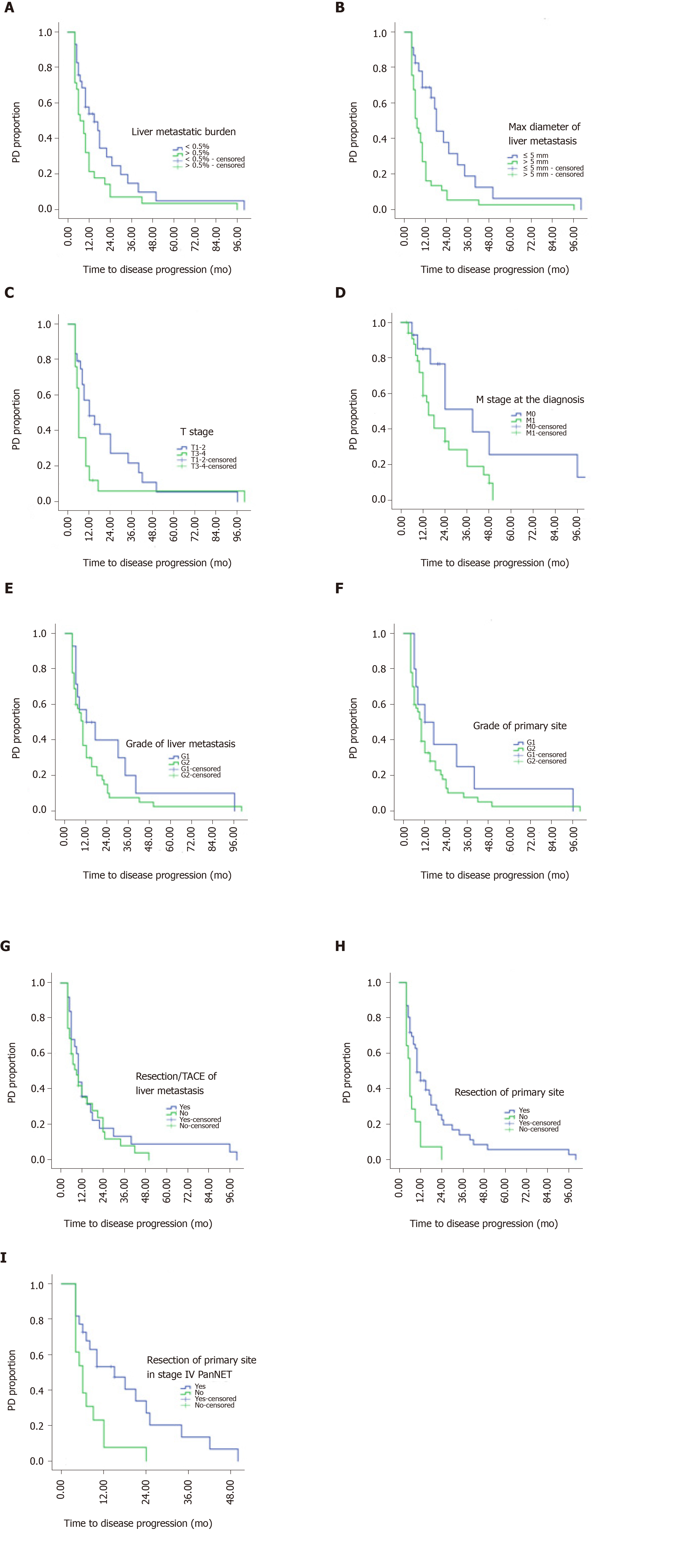
Figure 1 Determination of the clinicopathological factors predicting time to progression during surveillance of metastatic pancreatic neuroendocrine tumors.
A: Tumor burden of liver metastasis; B: Largest axis of the liver metastasis; C: T stage; D: M stage at diagnosis; E: Grade of liver metastasis; F: Grade of the primary tumor; G: Resection/transcatheter arterial chemoembolization of the liver metastasis; H: Resection of the primary tumor; I: Resection of the primary tumor in patients with stage IV disease. PanNET: Pancreatic neuroendocrine tumor; TACE: Transcatheter arterial chemoembolization; PD: Progressive disease.
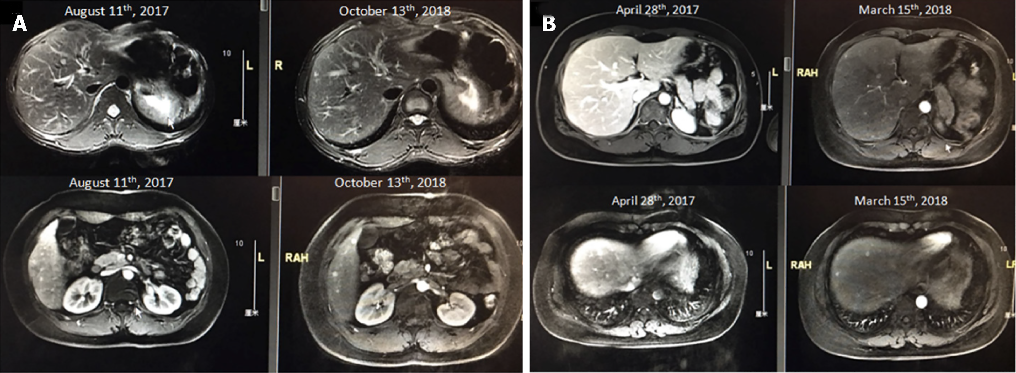
Figure 2 Magnetic resonance imaging screening showing an example of the largest axis of a liver metastasis < 5 mm.
A: Patient MRI scan image in 2017 August and 2018 October. B: Patient magnetic resonance imaging (MRI) scan image in 2017 April and 2018 March.
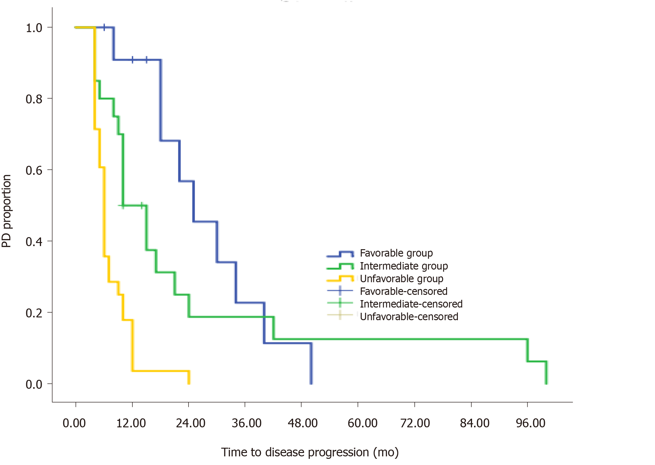
Figure 3 Risk stratification for predicting time to progression during surveillance on Kaplan-Meier survival curves of patients with metastatic pancreatic neuroendocrine tumors.
PD: Progressive disease.
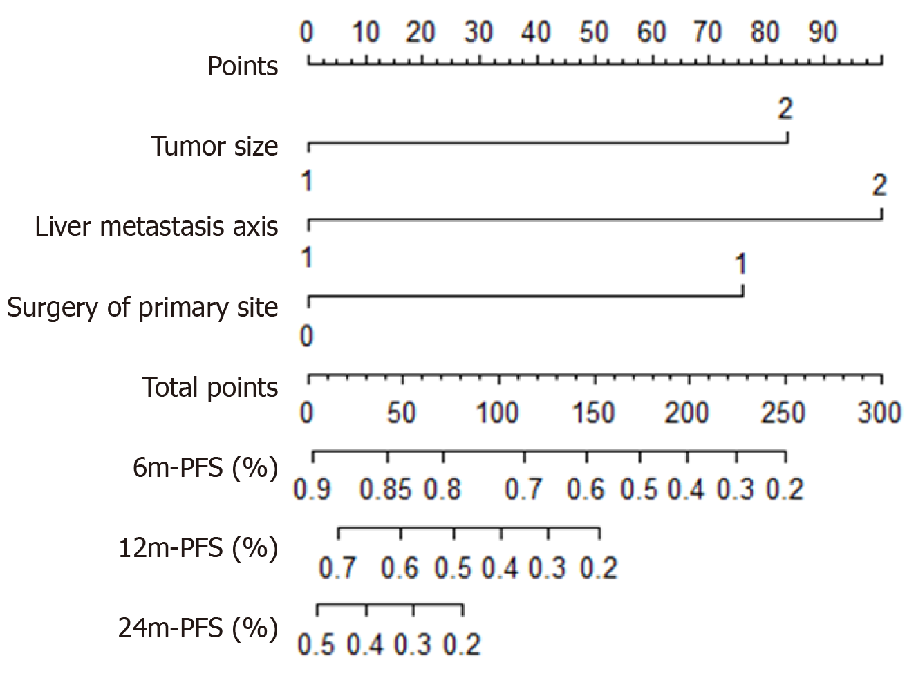
Figure 4 Nomogram for predicting time to progression during surveillance of metastatic pancreatic neuroendocrine tumors based on our proposed model.
Largest axis of the liver metastasis, T stage, and resection of the primary tumor. PFS: Progression-free survival.
- Citation: Gao HL, Wang WQ, Xu HX, Wu CT, Li H, Ni QX, Yu XJ, Liu L. Active surveillance in metastatic pancreatic neuroendocrine tumors: A 20-year single-institutional experience. World J Clin Cases 2020; 8(17): 3751-3762
- URL: https://www.wjgnet.com/2307-8960/full/v8/i17/3751.htm
- DOI: https://dx.doi.org/10.12998/wjcc.v8.i17.3751


