Copyright
©The Author(s) 2020.
World J Clin Cases. Aug 26, 2020; 8(16): 3542-3547
Published online Aug 26, 2020. doi: 10.12998/wjcc.v8.i16.3542
Published online Aug 26, 2020. doi: 10.12998/wjcc.v8.i16.3542
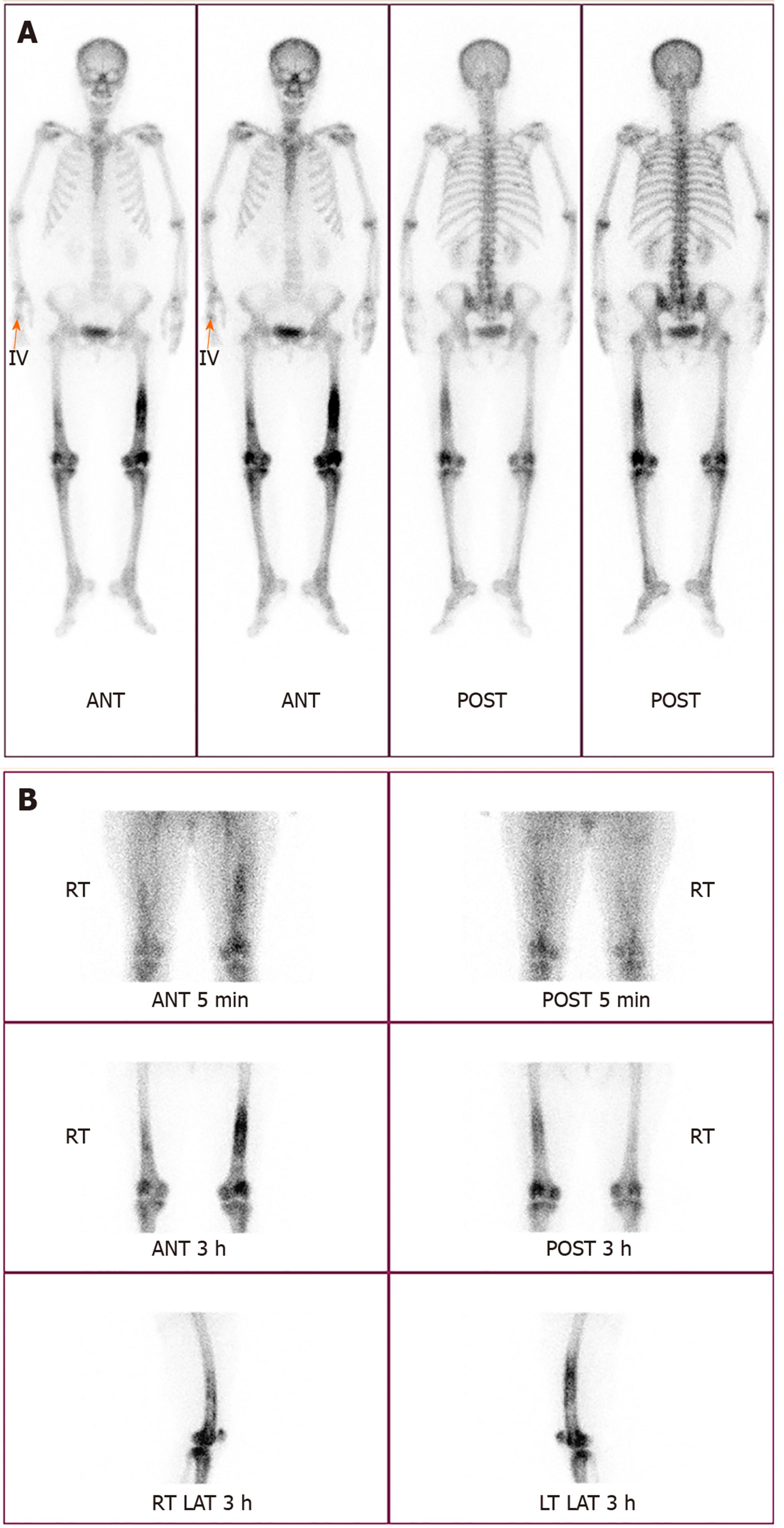
Figure 1 Tc-99m bone scan.
A: Diffuse increased uptake in the shaft of bilateral femurs (more severe in the left than on the right) and periarticular bones of bilateral knee joints; B: Blood pool phase (first row) revealing increased uptake in both femur shafts and knees; bone phase (second and third row) illustrates increased uptake in the same bones and joints.
Figure 2 Magnetic resonance imaging.
Diffuse irregular contours from a large intramedullary lesion are observed in both mid- to distal femurs (A), both proximal tibiae (B) and both patellae (C) (arrow head). There are T2 high and T1 low SI multiple fluid collection, from the diaphysis to the epiphysis and along the periosteum (arrow). These findings indicate osteomyelitis.
- Citation: Kim YJ, Lee JH. Disseminated osteomyelitis after urinary tract infection in immunocompetent adult: A case report. World J Clin Cases 2020; 8(16): 3542-3547
- URL: https://www.wjgnet.com/2307-8960/full/v8/i16/3542.htm
- DOI: https://dx.doi.org/10.12998/wjcc.v8.i16.3542


