Copyright
©The Author(s) 2020.
World J Clin Cases. May 26, 2020; 8(10): 2023-2027
Published online May 26, 2020. doi: 10.12998/wjcc.v8.i10.2023
Published online May 26, 2020. doi: 10.12998/wjcc.v8.i10.2023
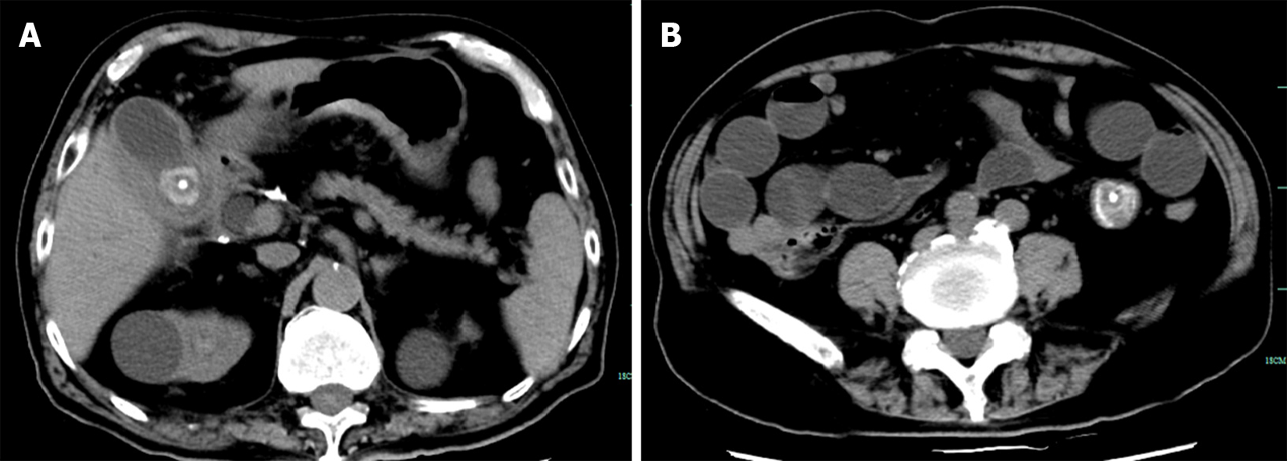
Figure 1 Imaging examinations.
A: Computed tomography image revealing cholecystolithiasis (approximately 2.6 cm in diameter); B: Circular high-density shadow in the intestinal cavity (approximately 3.5 cm in diameter).
Figure 2 Postoperative images.
A: On the fifth day after the operation, computed tomography (CT) examination revealed that cholecystolithiasis had disappeared according to the abdominal CT scan taken before the operation; B: The diameter of the stone in the bowel was approximately 2.0 cm, consistent with the shape of cholecystolithiasis on abdominal CT before the operation (Figure 1A); C: Gallstone was found in the small intestine during the second operation.
- Citation: Jiang H, Jin C, Mo JG, Wang LZ, Ma L, Wang KP. Rare recurrent gallstone ileus: A case report. World J Clin Cases 2020; 8(10): 2023-2027
- URL: https://www.wjgnet.com/2307-8960/full/v8/i10/2023.htm
- DOI: https://dx.doi.org/10.12998/wjcc.v8.i10.2023


