Copyright
©The Author(s) 2020.
World J Clin Cases. May 26, 2020; 8(10): 1979-1987
Published online May 26, 2020. doi: 10.12998/wjcc.v8.i10.1979
Published online May 26, 2020. doi: 10.12998/wjcc.v8.i10.1979
Figure 1 Radiologic and endoscopic features of the tumor.
A: Gastroscopy showed a nodular protruding lesion at the gastric antrum and stenosis of the pyloric ring; B: Abdominal computed tomography showed that the wall of the antrum was thickened and enhanced (arrow); C: Barium meal examination showed filling defect at the antrum and stenosis of the lumen.
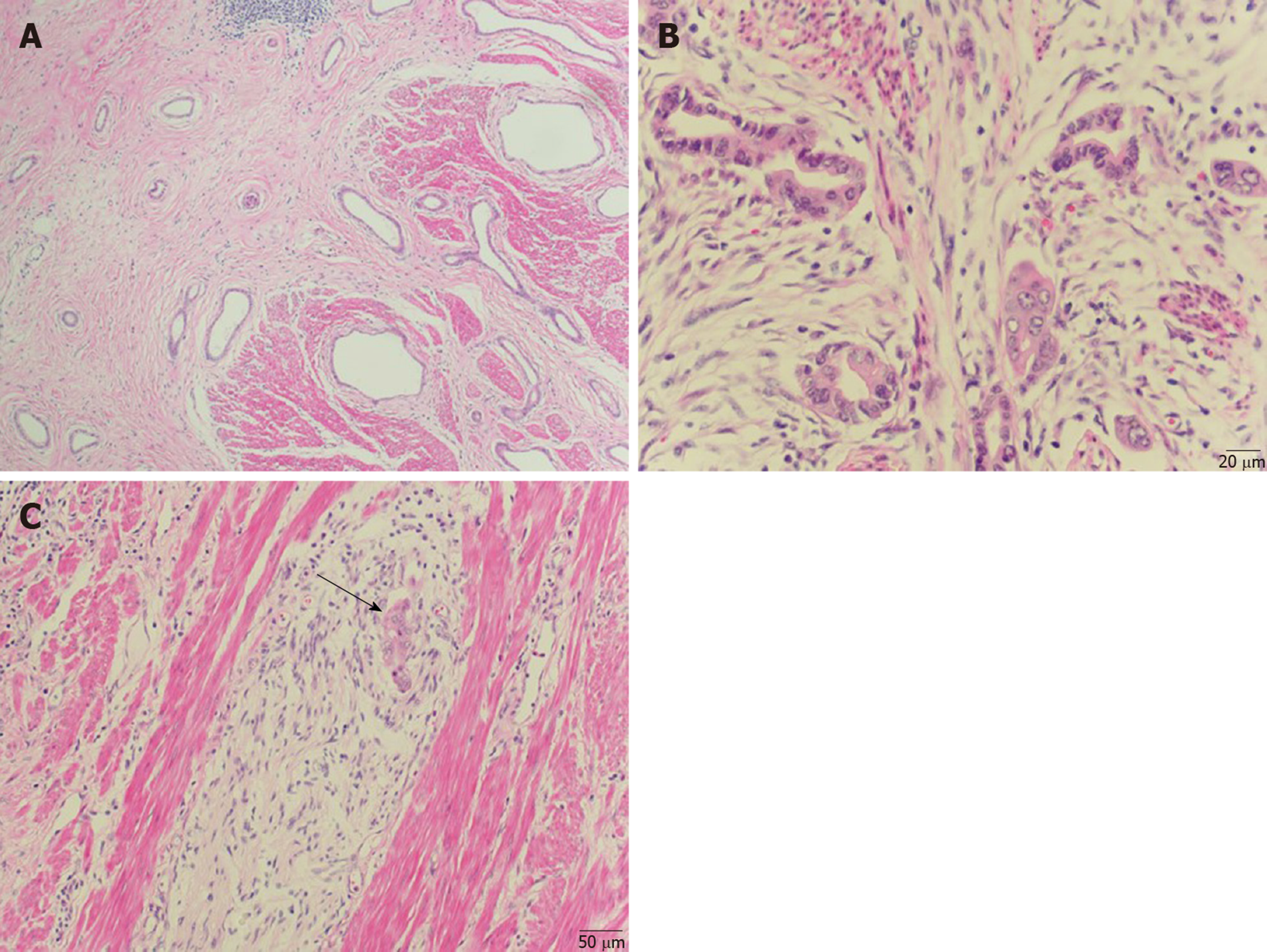
Figure 2 Hematoxylin-eosin histopathological examination of the surgically resected specimen.
A: A well-differentiated adenocarcinoma was revealed, coexisting with pancreatic heterotopia composed of ducts (×100); B: High-power view of duct-like structures of adenocarcinoma (×400); C: Invasion of bundles of nerves was seen (arrow) (×200).
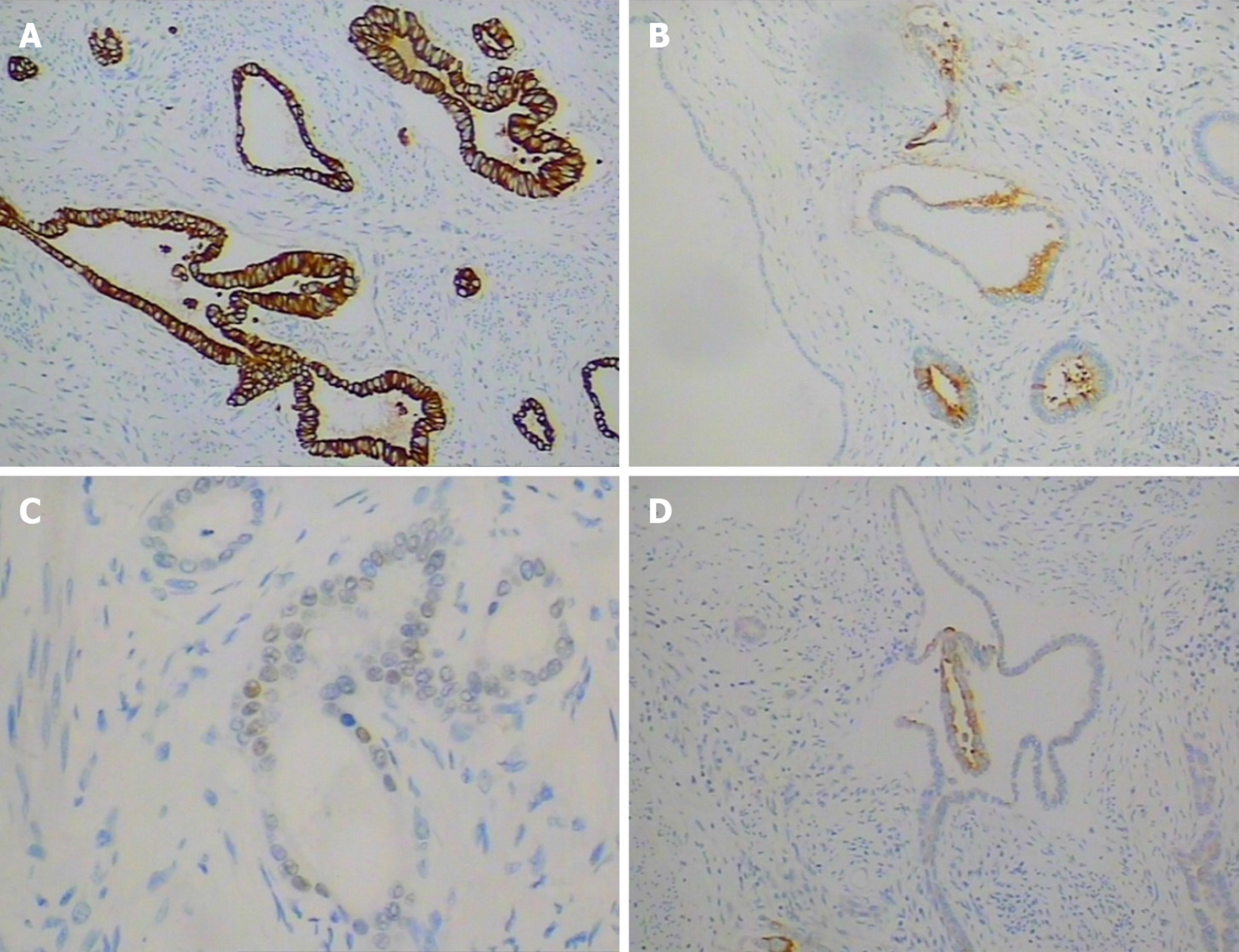
Figure 3 Immunohistochemical staining.
A: Cytokeratin 7 (×200); B: Carcinoembryonic antigen (×200); C: CDX-2 (×400); D: Cytokeratin 20 (×200).
- Citation: Xiong Y, Xie Y, Jin DD, Wang XY. Heterotopic pancreas adenocarcinoma in the stomach: A case report and literature review. World J Clin Cases 2020; 8(10): 1979-1987
- URL: https://www.wjgnet.com/2307-8960/full/v8/i10/1979.htm
- DOI: https://dx.doi.org/10.12998/wjcc.v8.i10.1979


