Copyright
©The Author(s) 2020.
World J Clin Cases. May 26, 2020; 8(10): 1973-1978
Published online May 26, 2020. doi: 10.12998/wjcc.v8.i10.1973
Published online May 26, 2020. doi: 10.12998/wjcc.v8.i10.1973
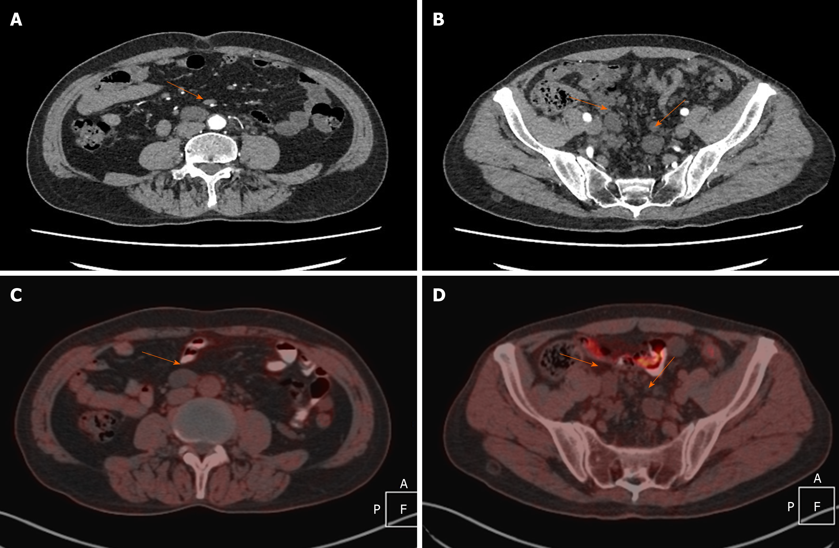
Figure 1 Contrast–enhanced abdominal-pelvic computed tomography and whole body positron emission tomography computed tomography images.
A and B: Axial contrast–enhanced computed tomography images showing multiple well-defined hypodense cystic lesions (orange arrows) arising from the retroperitoneum (A) and pelvic space (B) without enhancement. The lesions grew along the peritoneal interstitial space, the pelvic wall, and external iliac vascular deformation area, some of which showed shaping and suture-like changes. The biggest one was 3.3 cm × 2.5 cm. C and D: Positron emission tomography/computed tomography performed 1 h after injection of (10 mCi) 370 MBq of F-18-fluorodeoxyglucose showing that there was no uptake by the multiple retroperitoneum and pelvic masses.
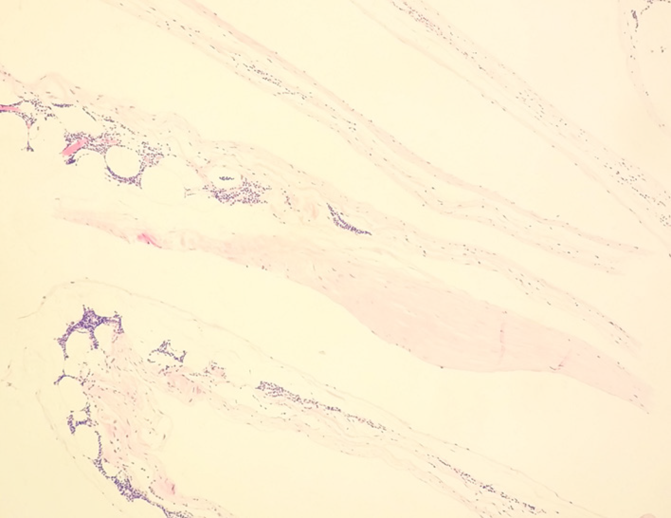
Figure 2 Fibrous cystic wall-like tissue observed under an optical microscope, with proliferated lymphoid tissue inside (HE staining, × 100).
The findings confirmed the lesions as multiple cystic lymphangiomas.
- Citation: Sun MM, Shen J. Positron emission tomography/computed tomography findings of multiple cystic lymphangiomas in an adult: A case report. World J Clin Cases 2020; 8(10): 1973-1978
- URL: https://www.wjgnet.com/2307-8960/full/v8/i10/1973.htm
- DOI: https://dx.doi.org/10.12998/wjcc.v8.i10.1973


