Copyright
©The Author(s) 2020.
World J Clin Cases. May 26, 2020; 8(10): 1958-1965
Published online May 26, 2020. doi: 10.12998/wjcc.v8.i10.1958
Published online May 26, 2020. doi: 10.12998/wjcc.v8.i10.1958
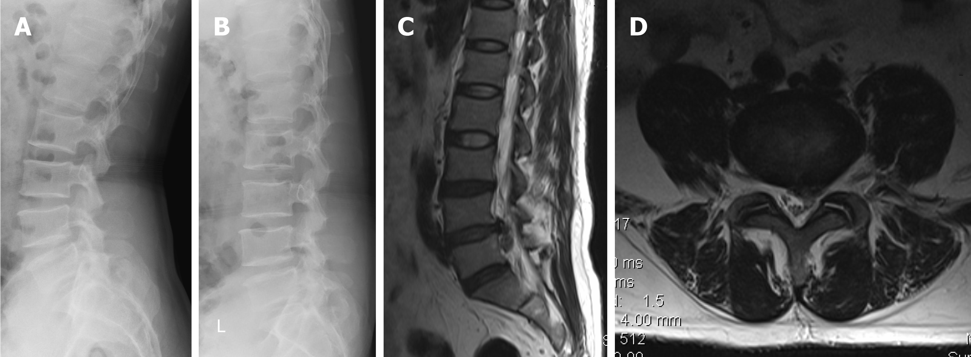
Figure 1 Preoperative imaging assessment.
A and B: Preoperative extension-flexion radiographs suggested decreased height without instability in the disc space (L4-5); C and D: Magnetic resonance imaging confirmed L3-S1 disc degeneration with associated low signal intensity and lumbar disc herniation (L4-5) on T2-weighted and axial images.
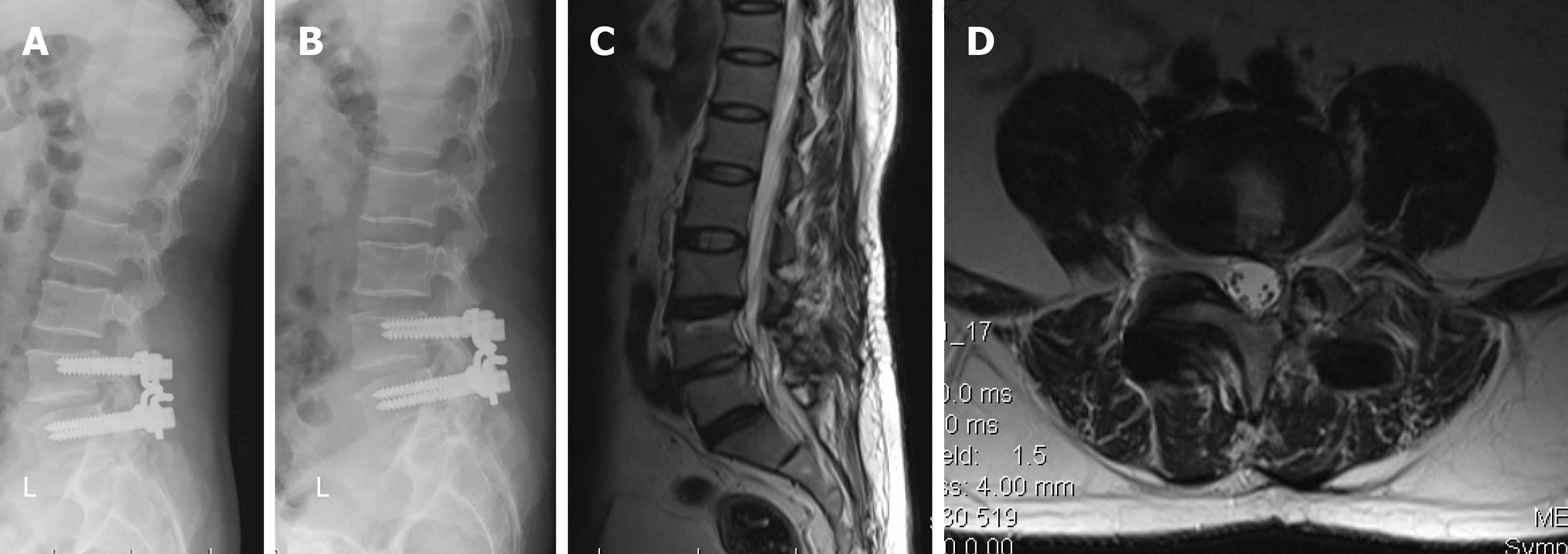
Figure 2 Postoperative imaging assessment.
A and B: Postoperative extension-flexion radiographs revealed that partial movement was reserved in the implanted level; C and D: Magnetic resonance imaging demonstrated disc rehydration within L4-5 at the 2-yr follow-up.
- Citation: Li YC, Feng XF, Pang XD, Tan J, Peng BG. Lumbar disc rehydration in the bridged segment using the BioFlex dynamic stabilization system: A case report and literature review. World J Clin Cases 2020; 8(10): 1958-1965
- URL: https://www.wjgnet.com/2307-8960/full/v8/i10/1958.htm
- DOI: https://dx.doi.org/10.12998/wjcc.v8.i10.1958


